Showing 17201–17250 of 36244 results
-

Intensifying screens 20 x 40 cm
$106.17 Add to cart View Product DetailsTo improve sensitivity and get sharp resolution. Made with calcium tungstenate. Highly suitable for molecular biology applications such as visualization of products labeled with P32, S35, I125.
-
Intensifying screens 24 x 30 cm
$294.85 Add to cart View Product DetailsTo improve sensitivity and get sharp resolution. Made with calcium tungstenate. Highly suitable for molecular biology applications such as visualization of products labeled with P32, S35, I125.
-
Intensifying screens 30 x 40 cm
$545.35 Add to cart View Product DetailsTo improve sensitivity and get sharp resolution. Made with calcium tungstenate. Highly suitable for molecular biology applications such as visualization of products labeled with P32, S35, I125.
-
Intensifying screens 35 x 43 cm
$642.56 Add to cart View Product DetailsTo improve sensitivity and get sharp resolution. Made with calcium tungstenate. Highly suitable for molecular biology applications such as visualization of products labeled with P32, S35, I125.
-
Interface cable RS232 for Titrette, length 2m, pack of 1
$194.81 Add to cart View Product DetailsInterface cable RS232 for Titrette, length 2m, pack of 1
-
Interleukin 2, human, recombinant
$600.05 Add to cart View Product DetailsInterleukin 2,Human, Recombinant
-
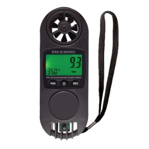
Intermediate Mini Environmental Quality Meter (8 Parameters)
$125.58 Add to cart View Product DetailsIntermediate Mini Environmental Quality Meter (8 Parameters)
-
Intrinsic factor
$132.68 Add to cart View Product DetailsIntrinsic Factor
-
Intrinsic factor
$262.91 Add to cart View Product DetailsIntrinsic Factor
-
Intrinsic factor
$865.72 Add to cart View Product DetailsIntrinsic Factor
-
Intrinsic factor
$1,731.61 Add to cart View Product DetailsIntrinsic Factor
1,505.75 -

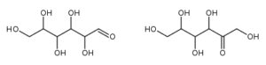
Inulin from chicory root
$46.24 Add to cart View Product DetailsInulin
-
Inulin from chicory root
$105.67 Add to cart View Product DetailsInulin
-
Inulin from chicory root
$342.49 Add to cart View Product DetailsInulin
-
Inulin from chicory root
$1,914.45 Add to cart View Product DetailsInulin
1,664.74 -
Invert sugar (pure syrup)
$41.22 Add to cart View Product DetailsInvert sugar (pure syrup)
-
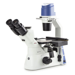
Inverted trinocular microscope with mech
$3,580.96 Add to cart View Product Detailsstage 10/20/40x,5W LED & w/transpor. box
-
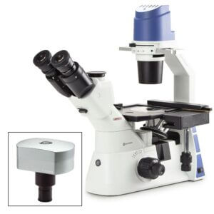
Inverted trinocular microscope with mech
$4,024.70 Add to cart View Product Detailsstage 10/20/40x, w/ camera
-
Invitrogen TrueGuide sgRNA Positive Control, AAVS1 (human), SKU: A35522
$299.28 Add to cart View Product DetailsProvides complete collection of validated positive controls for the TrueGuide Synthetic gRNA product line to help maximize editing performance. This positive control targeting AAVS1 targets intronic region of the AAVS1 locus and has achieved editing efficiencies of up to 60% in variety of human cell lines. AAVS1 controls easily determine the optimal conditions for delivering CRISPR/Cas9 tools to cell type, allowing to move forward in editing experiments and minimize downstream effort to isolate and validate your edited clones.
-

InvitrogenFGF1 Polyclonal Antibody, Biotin, PeproTech®
$449.85 Add to cart View Product DetailsnnAA Sequence of recombinant protein: MFNLPPGNYK KPKLLYCSNG GHFLRILPDG TVDGTRDRSD QHIQLQLSAE SVGEVYIKST ETGQYLAMDT DGLLYGSQTP NEECLFLERL EENHYNTYIS KKHAEKNWFV GLKKNGSCKR GPRTHYGQKA ILFLPLPVSS D.
Preparation: Produced from sera of rabbits immunized with highly pure Recombinant Human FGF-acidic. Anti-Human FGF-acidic-specific antibody was purified by affinity chromatography and then biotinylated.
Sandwich ELISA: To detect hFGF-acidic by sandwich ELISA (using 100 µL/well antibody solution) a concentration of 0.25-1.0 µg/mL of this antibody is required. This biotinylated polyclonal antibody, in conjunction with PeproTech Polyclonal Anti-Human FGF-acidic (500-P17) as a capture antibody, allows the detection of at least 0.2-0.4 ng/well of Recombinant hFGF-acidic.
Western Blot: To detect Human FGF-acidic by Western Blot analysis this antibody can be used at a concentration of 0.1-0.2 µg/mL. Used in conjunction with compatible secondary reagents the detection limit for Recombinant Human FGF-acidic is 1.5-3.0 ng/lane, under either reducing or non-reducing conditions.Target Information
FGF1 is a member of the fibroblast growth factor (FGF) family. FGF family members possess broad mitogenic and cell survival activities, and are involved in a variety of biological processes, including embryonic development, cell growth, morphogenesis, tissue repair, tumor growth and invasion. This protein functions as a modifier of endothelial cell migration and proliferation, as well as an angiogenic factor. It acts as a mitogen for a variety of mesoderm- and neuroectoderm-derived cells in vitro, thus is thought to be involved in organogenesis.
-
InvitrogenFGF1 Polyclonal Antibody, PeproTech®
$351.69 Add to cart View Product DetailsAA Sequence of recombinant protein: MFNLPPGNYK KPKLLYCSNG GHFLRILPDG TVDGTRDRSD QHIQLQLSAE SVGEVYIKST ETGQYLAMDT DGLLYGSQTP NEECLFLERL EENHYNTYIS KKHAEKNWFV GLKKNGSCKR GPRTHYGQKA ILFLPLPVSS D.
Preparation: Produced from sera of rabbits immunized with highly pure Recombinant Human FGF-acidic. Anti-Human FGF-acidic-specific antibody was purified by affinity chromatography employing an immobilized Human FGF-acidic matrix.
Sandwich ELISA: To detect Human FGF-acidic by sandwich ELISA (using 100 µL/well antibody solution) a concentration of 0.5-2.0 µg/mL of this antibody is required. This antigen affinity purified antibody, in conjunction with PeproTech Biotinylated Anti-Human FGF-acidic (500-P17Bt) as a detection antibody, allows the detection of at least 0.2-0.4 ng/well of Recombinant Human FGF-acidic.
Western Blot: To detect Human FGF-acidic by Western Blot analysis this antibody can be used at a concentration of 0.1-0.2 µg/mL. When used in conjunction with compatible secondary reagents, the detection limit for Recombinant Human FGF-acidic is 1.5-3.0 ng/lane, under either reducing or non-reducing conditions. -
Invitrogen™ BODIPY™ TR Ceramide SKU: d7540
$401.35 Add to cart View Product DetailsBeschreibung
BODIPY TR ceramide can be used to stain Golgi apparatus in live cells.
Cell Analysis, Cell Structure, Golgi
Bestellinformation
Shipping Condition: Room Temperature
-
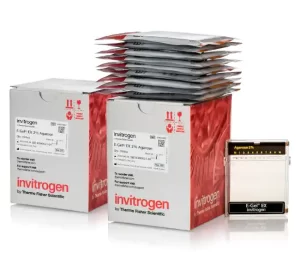
Invitrogen™ E-Gel™ EX Agarose Gels, 2%
$397.90 Add to cart View Product Details-
- Contain a proprietary fluorescent nucleic acid stain (excitation: 490nm, emission: 520nm) that can be viewed with blue light, which minimizes DNA damage and allows detection down to 1ng/band of DNA
n
-
- Complete separation (by molecular weight) in 10 minutes
n
-
- Live monitoring of DNA migration without the hazards of UV light
n
-
- Easy access to the gel for extraction of DNA bands or transfer to membranes for Southern blotting
n
-
- Demonstrate over five-fold greater sensitivity than comparable gels containing ethidium bromide allows less sample use per run
n
-
- Fast analysis of RNA samples to check on their integrity before proceeding with downstream application
n
-
- The gel cassette is designed to open with a Gel Knife, so that the gel inside can be accessed readily for excision of specific bands or transferred to a membrane for Southern blot analysis
n
-
- Single comb well format
n
-
- Sample Type: Double-Stranded DNA (dsDNA), Single-Stranded RNA
n
-
- Sample Volume; 20µL
n
Agarose Gel Electrophoresis, DNA and RNA Purification and Analysis, Nucleic Acid Gel Electrophoresis and Blotting
Order Info
Shipping Condition: Room Temperature
-
-
Invitrogen™ LV-MAX™ Transfektionskit SKU: a35348
$1,897.50 Add to cart View Product DetailsThe Gibco LV- MAX Transfection Kit is an essential component of the LV-MAX Lentiviral Production System. It is formulated for the transient transfection of Gibco Viral Production Cells (high-density suspension-adapted human embryonic kidney (HEK) 293-derived cells). The included high-efficiency, cationic, lipid-based transfection reagent, lentiviral production supplement, and LV enhancer are designed to work together to produce the highest possible level of lentiviral particles from the Viral Production Cells when cultured in LV-MAX Production Medium.
-
- Designed specifically for transfection of high-density suspension cells to maximize lentiviral production
n
-
- Enables sustained, high-level production of high-density transiently-transfected cultures, achieving titer of up to 1 x 10⁸ TU/mL (unconcentrated LVVs-GFP)
n
-
- Reproducible and scalable transfection results, giving users greater confidence in their results
n
-
- Easy, robust culture and transfection protocols—fits into any transient production workflows.The kit includes three key components required to optimize your lentiviral production. These are only available in the kit and are not sold separately.
n
-
- LV-MAX transfection reagent—a high-efficiency, cationic, lipid-based transfection reagent uniquely designed for co-transfecting multiple plasmids into high-density Viral Production Cell cultures with high transfection efficiency and low toxicity
n
-
- LV-MAX supplement—an optimized, chemically defined, serum-free, protein-free, animal origin-free formulation designed to suppress cell growth during transfection and increase lentiviral vector production without compromising the cell viability and interfering with the lentiviral production process
n
-
- LV-MAX enhancer—a proprietary, animal origin-free formulation designed to boost cell lentiviral vector production
n
-
-
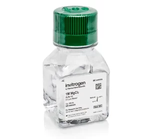
Invitrogen™ MgCl2 (1 M)
$78.97 Add to cart View Product DetailsMolecular biology grade, Invitrogen 1M MgCl2 solution is supplied in one bottle containing 100mL. The solution is certified RNase-free, economical, and ready-to-use. Due to the ubiquitous presence of RNases, manufacturing products for use with RNA is especially challenging. Invitrogen’s nuclease-free reagents and buffers are manufactured in facilities specifically designed to prevent the introduction of nucleases. Highly sensitive RNase assays are performed at several different stages of the manufacturing process to ensure the highest quality. These reagents are rigorously tested for contaminating nonspecific endonuclease, exonuclease, and RNase activity.n
Order Info
Shipping Condition: Room temperature
-
Invitrogen™ Neon™ Transfection System 100 ?L Kit
$3,055.12 Add to cart View Product Details-
- For use with the Neon Transfection System
n
-
- Use this kit for transfection volumes of 100µL, containing 5 x 10⁵-2 x 10⁶ adherent cells or 1 x 10⁶-5 x 10⁶ suspension cells
n
-
- Cells that have been transfected using the included 100µL Neon Tips are then ready to be washed and plated into a 6-well, 60mm, or 10cm culture dish, depending on protocol
n
Adenoviral shRNA, Cell Culture, Lentiviral shRNA & miRNA, Plasmid Transfection, RNAi, RNAi Transfection, RNAi, Epigenetics & Noncoding RNA Research, Retroviral RNAi, Stem Cell & Primary Cell Transfections, Synthetic siRNA Transfection, Transfection, Vector-Based RNAi, siRNA
Order Info
Shipping Condition: Room Temperature
-
-
Invitrogen™ Novex™ Tris-Glycine Mini Protein Gels, 10–20%, 1.0 mm, WedgeWell™ format X
$154.10 Add to cart View Product DetailsInvitrogen Novex Tris-Glycine Mini Gels, WedgeWell format, are polyacrylamide gels that provide reproducible separation of a wide range of proteins into well-resolved bands with two times the loading capacity. Novex Tris-Glycine Mini Gels are based on traditional Laemmli protein electrophoresis that allow the use of Laemmli sample and running buffers.
nFeatures of Novex Tris-Glycine gels, WedgeWell format, include:
• Improved shelf life—store gels for up to 21 weeks at 4°C
• Diversity—use for native and denaturing protein assays
• Wedge-shaped wells—easily load up to two times more sample volume
• Fast run conditions—quickly separate your proteins using constant voltage in less than 60 minutes
Choose the right gel format for your experiments
Novex Tris-Glycine gels, WedgeWell format, come in a variety of fixed concentrations from 6% to 18%, as well as gradients with ranges of 4–12%, 4–20%, 8–16%, and 10–20%. You can select from our many well formats, including 10-, 12-, and 15-well, and different pack sizes.
Run your proteins in native or denatured form
Novex Tris-Glycine gels, WedgeWell format, do not contain SDS and can be used to run your proteins in native or in denatured form. For denatured proteins, we recommend using Novex Tris-Glycine SDS Sample Buffer (LC2676) and Novex Tris-Glycine SDS Running Buffer (LC26755). For native proteins, we recommend using Novex Tris-Glycine Native Sample Buffer (LC2673) and Novex Tris-Glycine Native Running Buffer (LC2672). -
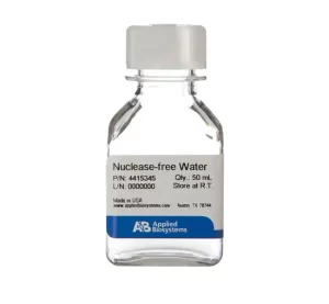
Invitrogen™ Nuclease-Free Water (not DEPC-Treated)
$210.60 Add to cart View Product Details-
- Nuclease-free Water (not DEPC-treated) has been deionized, filtered into the final bottle, and autoclaved
n
-
- It is ready to use and requires no preparation, mixing, or autoclaving
n
-
- Rigorously tested for contaminating nonspecific endonuclease, exonuclease, and RNase activity
n
-
- Since this water is not DEPC-treated, it is recommended for applications in which residual DEPC might prove detrimental (e.g., oocyte microinjection)
n
-
-
Invitrogen™ NuPAGE™ 4 to 12%, Bis-Tris, 1.0 mm, Midi Protein Gel, 26-well SKU: wg1403box
$258.75 Add to cart View Product DetailsInvitrogen NuPAGE Bis-Tris protein gels are precast polyacrylamide gels that provide broad molecular weight protein separation with high resolution and sample integrity. These precast gels are ideal for applications where protein integrity is crucial. Unlike traditional Tris-glycine gels, NuPAGE Bis-Tris gels have a neutral pH environment that minimizes protein modifications. Use NuPAGE Bis-Tris gels for preparing proteins for sequencing, mass spectrometry, and any other techniques where protein integrity is important.
nFeatures of NuPAGE Bis-Tris gels:
• Better protein integrity—optimized sample preparation process preserves your proteins
• Wide ranges of molecular weight separation—select the right gel and running buffer to get the optimal separation of your proteins
• Faster run times—get separation of your proteins in as little as 35 minutes
• Longer shelf life—NuPAGE Bis-Tris gels can be stored for at least 12 months at room temperature
Choose the right NuPAGE Bis-Tris gel for your protein separation
Obtain optimal separation of your proteins by choosing the right combination of gel and running buffer. NuPAGE Bis-Tris protein gels come in four polyacrylamide concentrations: 8%, 10%, 12%, and a 4–12% gradient. Gels come in two sizes: mini (8 cm x 8 cm) or midi (8.7 cm x 13.3 cm) and either 1.0 mm (mini and midi gels) or 1.5 mm (mini gel format only) in thickness. NuPAGE Bis-Tris gels also come in multiple well formats.
NuPAGE Bis-Tris gels are formulated for denaturing gel electrophoresis applications. For optimal sample preparation, use the NuPAGE LDS Sample Buffer (NP0007) and NuPAGE Sample Reducing Agent (NP0004). Use NuPAGE Antioxidant (NP0005) in the running buffer to maintain the reduced state of the proteins during the run and to allow maximum band sharpness. The gels can be run using NuPAGE MES SDS Running Buffer (NP0002) to better resolve small proteins or NuPAGE MOPS SDS Running Buffer (NP000102) to resolve medium- to large-size proteins.
NuPAGE Bis-Tris Midi gels can be used with the XCell SureLock Midi Cell gel apparatuses (WR0100) or conveniently with the Bio-Rad Criterion Cell using our adapters (WA0999).
For transfer of proteins to a membrane, rapid semi-dry transfer can be done using the Invitrogen Power Blotter or rapid dry transfer using the iBlot 2 Gel Transfer Device (IB21001). Alternatively, traditional wet transfer can be performed using the Bio-Rad Criterion Cell using NuPAGE Transfer buffer (NP00061). -
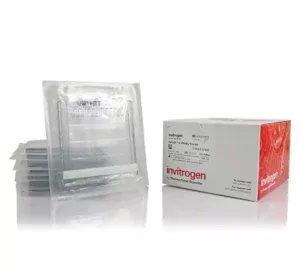
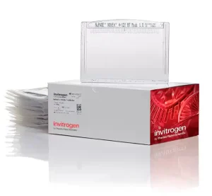
Invitrogen™ NuPAGE™ 4 to 12%, Bis-Tris, 1.0–1.5 mm, Mini Protein Gels
$224.46 Add to cart View Product DetailsInvitrogen NuPAGE Bis-Tris protein gels are precast polyacrylamide gels that provide broad molecular weight protein separation with high resolution and sample integrity. These precast gels are ideal for applications where protein integrity is crucial. Unlike traditional Tris-glycine gels, NuPAGE Bis-Tris gels have a neutral pH environment that minimizes protein modifications. Use NuPAGE Bis-Tris gels for preparing proteins for sequencing, mass spectrometry, and any other techniques where protein integrity is important.
Features of NuPAGE Bis-Tris gels:
• Better protein integrity—optimized sample preparation process preserves your proteins
• Wide ranges of molecular weight separation—select the right gel and running buffer to get the optimal separation of your proteins
• Faster run times—get separation of your proteins in as little as 35 minutes
• Longer shelf life—NuPAGE Bis-Tris gels can be stored for at least 12 months at room temperature
Choose the right NuPAGE Bis-Tris gel for your protein separation
Obtain optimal separation of your proteins by choosing the right combination of gel and running buffer. NuPAGE Bis-Tris protein gels come in four polyacrylamide concentrations: 8%, 10%, 12%, and a 4–12% gradient. Gels come in two sizes: mini (8 cm x 8 cm) or midi (8.7 cm x 13.3 cm) and either 1.0 mm (mini and midi gels) or 1.5 mm (mini gel format only) in thickness. NuPAGE Bis-Tris gels also come in multiple well formats.
NuPAGE Bis-Tris gels are formulated for denaturing gel electrophoresis applications. For optimal sample preparation, use the NuPAGE LDS Sample Buffer (NP0007) and NuPAGE Sample Reducing Agent (NP0004). Use NuPAGE Antioxidant (NP0005) in the running buffer to maintain the reduced state of the proteins during the run and to allow maximum band sharpness. The gels can be run using NuPAGE MES SDS Running Buffer (NP0002) to better resolve small proteins or NuPAGE MOPS SDS Running Buffer (NP000102) to resolve medium- to large-size proteins.
For transfer of proteins to a membrane, we recommend using NuPAGE Transfer Buffer (NP00061) for traditional wet transfer using the Mini Blot Module (B1000) or XCell II Blot Module (EI9051). Alternatively, rapid semi-dry transfer can be done using the Invitrogen Power Blotter or rapid dry transfer using the iBlot 2 Gel Transfer Device (IB21001). -
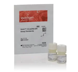
Invitrogen™ Qubit™ 1X dsDNA Assay Kits, high sensitivity (HS) and broad range (BR) SKU: q33266
$455.00 Add to cart View Product DetailsExperience easy, accurate, and precise quantification of dsDNA with Qubit 1X dsDNA HS (High Sensitivity) and Qubit 1X dsDNA BR (Broad Range) Assay Kits. The 1X formulation provides a ready-to-use working solution that enables simple dsDNA sample quantification on Qubit Fluorometers. These assays are highly selective for dsDNA over ssDNA, RNA, protein, and free nucleotides. Contaminants, such as salts, solvents, or detergents are well-tolerated.
nQubit 1X dsDNA HS and BR Assay Kits, designed for use with Qubit Fluorometers, are highly selective for double-stranded DNA (dsDNA) over single-stranded DNA (ssDNA), RNA, protein, and free nucleotides. All kits provide a ready-to-use working solution and DNA standards. To perform the assay, simply dilute your sample (any volume from 1–20 µL is acceptable) into the 1X working solution provided, then read the concentration using a Qubit Fluorometer.
Qubit 1X dsDNA HS Assay Kit
The Qubit dsDNA HS (High Sensitivity) Assay Kit, when used with the Qubit Fluorometer, provides an accurate and selective method for the quantitation of sensitive DNA samples. Depending on sample volume, the assay kit is designed to be accurate for initial DNA sample concentrations of 5 pg/µL to 120 ng/µL, providing a detection range of 0.1-120 ng.
Qubit 1X dsDNA BR Assay Kit
The Qubit dsDNA BR (Broad-Range) Assay Kit, designed for use with Qubit 4 or Qubit Flex Fluorometers, provides an accurate and selective method for the quantitation of DNA samples. Depending on sample volume, the assay kit is designed to be accurate for initial DNA sample concentrations of 0.2 to 4,000 ng/µL, providing a detection range of 4-4,000 ng.
Notes
• Qubit 1X dsDNA HS can be used with Qubit 2, Qubit 3, Qubit 4, and Qubit Flex Fluorometers
• Qubit 1X dsDNA BR are designed for use with Qubit 4 and Qubit Flex Fluorometers
• Use thin-wall, clear, 0.5-mL PCR tubes (Cat. No. Q32856) for the Qubit 4 Fluorometer and 8 x 200 µL tube strips (Cat. No. Q33252) for the Qubit Flex Fluorometer -
Invitrogen™ Qubit™ Assay Tubes
$159.33 Add to cart View Product Details-
- Qubit assay tubes are 500µL, thin-walled polypropylene tubes for use with the Qubit Fluorometer
n
-
- 500 tubes per package
n
Order Info
Shipping Condition: Room temperature
-
-
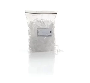
Invitrogen™ Qubit™ Flex Assay Tube Strips SKU: q33252
$351.92 Add to cart View Product DetailsQubit™ Flex Assay Tube Strips are 200-µL thin-walled polypropylene tubes in a strip of eight tubes that are for use with the Qubit™ Flex Fluorometer.
- Capacity: 200 ?L
- Thin-walled
- Polypropylene tubes
- For use with the Qubit™ Flex Fluorometer
- Quantity: 125 tube strips
- Strips of eight tubes
-
Invitrogen™ Qubit™ RNA high sensitivity (HS), broad range (BR) and extended range (XR) Assay Kits SKU: q32852
$178.73 Add to cart View Product DetailsAchieve accurate and precise quantification of RNA with Qubit RNA HS, BR, and XR Assay Kits. These RNA quantification kits enable quick and selective detection of low and high abundance RNA samples, and can distinguish RNA from DNA, protein, and free nucleotides. Contaminants such as salts, solvents, or detergents are well-tolerated.
nQubit RNA HS, BR, and XR Assay Kits, designed for use with Qubit Fluorometers, are highly selective for RNA over DNA, protein, and free nucleotides. All kits provide concentrated assay reagent, dilution buffer, and pre-diluted RNA standards. Simply dilute the reagent using the buffer provided, add your sample (any volume between 1 µL and 20 µL is acceptable), and read the concentration using a Qubit Fluorometer.
Qubit RNA HS Assay Kit
The Qubit RNA HS (High Sensitivity) Assay Kit, when used with the Qubit Fluorometer, provides an accurate and selective method for the quantitation of low-abundance RNA samples. Depending on sample volume, the assay kit is designed to be accurate for initial RNA sample concentrations of 0.2 to 200 ng/µL, providing a detection range of 4-200 ng.
Qubit RNA BR Assay Kit
The Qubit RNA BR (Broad-Range) Assay Kit, when used with the Qubit Fluorometer, provides an accurate and selective method for the quantitation of RNA samples. Depending on sample volume, the assay kit is designed to be accurate for initial RNA sample concentrations of 0.5 to 1,200 ng/µL, providing a detection range of 10-1,200 ng.
Qubit RNA XR Assay Kit
The Qubit RNA XR (Extended Range) Assay Kit, designed for use with Qubit 4 and Qubit Flex Fluorometers, provides an accurate and selective method for the quantitation of high-abundance RNA samples. Depending on sample volume, the assay kit is designed to be accurate for initial RNA sample concentrations of 5 to 20,000 ng/µL, providing a detection range of 100-20,000 ng.
Notes
• Qubit RNA HS and BR Assay kits can be used with any Qubit Fluorometer
• Qubit RNA XR Assay Kits are designed to be used only with Qubit 4 or Qubit Flex Fluorometers
• Use with thin-wall, clear, 0.5-mL PCR tubes (Cat. No. Q32856) for the Qubit 4 Fluorometer and 8 x 200-µL tube strips (Cat. No. Q33252) for the Qubit Flex FluorometerOrder Info
Shipping Condition: Room temperature
-
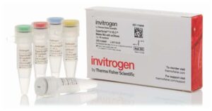
Invitrogen™ SuperScript™ IV VILO™ Master Mix X
$1,022.53 Add to cart View Product DetailsSuperScript™ IV VILO™ (SSIV VILO) Master Mix is a reaction master mix designed for fast, sensitive, and reproducible cDNA synthesis in RT-qPCR applications. Inclusion of ezDNase, further accelerates the RT-qPCR workflow through an extremely simplified genomic DNA removal step. This master mix is also available without ezDNase, which can be purchased separately.
Features of SuperScript IV VILO Master Mix include:n-
- Super-fast RT reaction in just 10 minutes and gDNA removal in 2 minutes
n
-
- Super-high yields—lower Ct values by more than 2 cycles ahead of all other reverse transcription reagents
n
-
- Super-convenient one-tube cDNA reaction master mix for two-step RT-qPCR
n
-
- Super-efficient reaction even with low template amounts and suboptimal purity samples
n
SSIV VILO master mix elevates the trusted VILO technology to the next level with the highly processive and thermostable SuperScript™ IV Reverse Transcriptase and further optimized buffer. These components enable efficient cDNA synthesis at higher temperatures and in less time. SSIV VILO master mix provides superior cDNA yield and sensitivity even with suboptimal purity or scarce templates. It is your new tool for more efficient and reproducible RT-qPCR.
For even better performance, some SuperScript IV VILO Master Mix products include ezDNase (also available as a separate purchase) for genomic DNA removal to further accelerate the RT-qPCR workflow. The extremely simplified genomic DNA removal step dramatically reduces the time of the entire reverse transcription protocol and reduces possible variation in gene expression due to RNA loss or damage during conventional DNase treatment.
SSIV VILO master mix gives you more cDNA in less time with less pipeting, less variation, less reaction inhibition, less gDNA interference, and with less sample than all the other cDNA synthesis kits or master mixes for RT-qPCR.
Source
Purified from E. coli expressing the pol gene of M-MLV, modified to increase thermostability and half-life, processivity, inhibitor resistance, and to reduce RNase H activity.
Performance and quality testing
Assayed for endodeoxyribonuclease, exodeoxyribonuclease, and ribonuclease, as well as yield and length of cDNA product. -
-
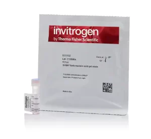
Invitrogen™ SYBR™ Safe DNA Gel Stain SKU: s33102
$133.71 Add to cart View Product DetailsSYBR Safe stain is specifically formulated to be a less hazardous alternative to ethidium bromide that can utilize either blue light or UV excitation.
-
- Reduces exposure to highly mutagenic ethidium bromide and harmful UV light
n
-
- Increases sensitivity by reducing nonspecific background fluorescence
n
-
- Use in place of ethidium bromide for all staining applications, including RNA staining
n
-
- Less hazardous
n
-
-
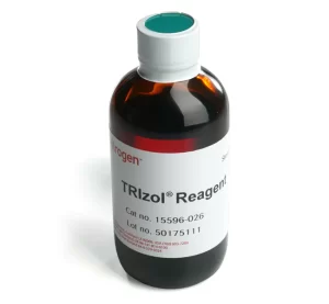
Invitrogen™ TRIzol™ Reagent- SKU: 15596026
$339.46 Add to cart View Product DetailsTRIzol™ Reagent is a complete, ready-to-use reagent for the isolation of high-quality total RNA or the simultaneous isolation of RNA, DNA, and protein from a variety of biological samples. This monophasic solution of phenol and guanidine isothiocyanate is designed to isolate separate fractions of RNA, DNA, and proteins from cell and tissue samples of human, animal, plant, yeast, or bacterial origin, within one hour.
-
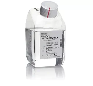
Invitrogen™ UltraPure™ 1 M Tris-HCI Buffer, pH 7.5
$94.22 Add to cart View Product Details-
- Prepared as 1M concentrate, buffer can be diluted to desired concentration
n
-
- Used in molecular biology or general biochemistry applications
n
-
- Performance and quality testing: No DNase, RNase, or protease activity detected
n
-
- Sustainable packaging
n
Agarose Gel Electrophoresis, DNA & RNA Purification & Analysis, DNA Extraction, General gDNA Purification Reagents & Accessories, Genomic DNA Purification, Nucleic Acid Gel Electrophoresis & Blotting
Order Info
Shipping Condition: Room Temperature
-
-
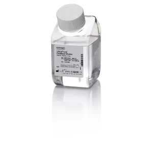
Invitrogen™ UltraPure™ DNase/RNase-Free Distilled Water
$67.47 Add to cart View Product Details-
- UltraPure DNase/RNase-Free Distilled Water is 0.1-µm membrane-filtered and tested for DNase and RNase activity
n
-
- Performance and quality testing: No DNase, RNase, or protease activity detected. Our distilled water system is routinely monitored for compliance with current USP monograph test requirements for Water for Injection (WFI)
n
-
- Sustainable packaging
n
-
-

Invitrogen™ ZOOM™ IPGRunner™ Cassettes and Trays SKU: zm0008
$696.50 Add to cart View Product DetailsZOOM Equilibration Trays are designed for use with the ZOOM IPGRunner Cassettes to perform equilibration of ZOOM Strips after first-dimension isoelectric focusing and prior to second-dimension (2D) SDS-PAGE.
-
- Simplify the equilibration and incubation process in 2D electrophoresis
n
-
- Savings in time and labor
n
-
- Optimal equilibration of samples
n
-
- Minimal risk of sample contamination and damage to IPG strips
n
Gel Electrophoresis, IsoElectric Focusing, Protein Gel Electrophoresis, Protein Sample Fractionation, Protein Sample Preparation and Protein Purification, Proteins, Expression, Isolation and Analysis
-
-
Invitrogen™Cell Culture Contamination Detection Kit
$429.47 Add to cart View Product DetailsThe Cell Culture Contamination Detection Kit is a simple and effective procedure for screening tissue cultures for contamination by microorganisms. The kit not only detects contaminants, but it also identifies the contaminant type. Three fluorescent dyes distinctly stain yeast (and other fungi), gram-positive and gram negative bacteria by slide preparation.nnBioproduction, Cell Analysis, Cell Culture, Cell Tracing and Tracking, Clinical, Contaminant and Impurity QC Testing, Infectious Disease Diagnostics, Mammalian Cell Culture, Microbial Tracking, Molecular Diagnostics, Mycoplasma Detection
-
Invitrogen™Cells-to-CT™ Bulk Fast Advanced RT Reagents X
$13,176.50 Add to cart View Product DetailsInvitrogen Fast Advanced Cells-to-CT Bulk RT Reagents are separately-available, large-quantity versions of the reagents used for reverse transcription (RT) in the TaqMan Fast Advanced Cells-to-CT Kit and the SYBR Green Fast Advanced Cells-to-CT Kit (20X RT enzyme mix and 20X RT buffer).
-
Invitrogen™CellsDirect Resuspension & Lysis Buffers includes 10 ml Resuspension Buffer & 1 ml Lysis Buffer X
$1,684.31 Add to cart View Product DetailsThese two buffers are to be used only as a complement to the SuperScript™ III Platinum™ CellsDirect Kits when large number of samples are processed. See manual of SuperScript III Platinum CellsDirect Kits for protocol recommendations.
-
- Fewer resources used, less hazardous, less waste
PCR and Real-Time PCR, Real Time PCR (qPCR), Real-Time PCR Directly from Samples
-
-
Invitrogen™GeneArt™ Genomic Cleavage Detection Kit
$440.54 Add to cart View Product DetailsThe GeneArt Genomic Cleavage Detection Kit provides a simple, reliable, and rapid method for the detection of locus-specific double-strand break formation. When using genome editing tools to obtain targeted mutations, it is necessary to determine the efficiency at which these nucleases cleave the target sequence, particularly prior to proceeding to the more laborious and expensive processes of cloning and sequencing.nnThe GeneArt genomic cleavage detection kit contains all of the reagents required prior to gel analysis. A sample of the edited cell population is used as a direct PCR template for amplification with primers specific to the targeted region. The PCR product is then denatured and re-annealed to produce heteroduplex mismatches where double strand breaks have occurred, resulting in indel introduction. These mismatches are recognized and cleaved by the detection enzyme. This cleavage is both easily detectable and quantifiable using gel analysis.
-
Invitrogen™SYBR™ Green Fast Advanced Cells-to-CT™ Kit X
$824.15 Add to cart View Product DetailsThe SYBR Green Fast Advanced Cells-to-Ct Kit is a complete cell lysate system designed for gene expression analysis directly from cultured cells without RNA purification. SYBR Green Fast Advanced Cells-to-Ct cell lysis reagents are supplied with highest sensitivity Cells-to-Ct Fast Advanced reverse transcription enzyme mix, 2X RT buffer, and best-in-class PowerUp SYBR Green Master mix. The SYBR Green Fast Advanced Cells-to-Ct Kit gives you the sensitivity you need in the fastest workflow available: get gene expressions analysis results from an entire plate of cells in about 1.5 hours.
• Complete—optimized workflow includes cell lysis reagents with gDNA removal, new Fast Advanced RT enzyme mix, buffer, and new PowerUp SYBR Green Fast Advanced Master Mix
• Fast—7-minute sample prep, including DNase treatment, at room temperature
• Easy—lyse samples in a tube or directly in culture plates
• Robust—perform gene expression analysis on 10–100,000 cells per sample; results equivalent to those from purified RNA
• Efficient—contains sufficient reagents to generate 1000 real-time PCR results from 100 starting samples
Unique method eliminates the need to isolate RNA from cultured cells
The Cells-to-Ct method features a unique lysis reagent that lyses cells and protects RNA. DNA removal occurs simultaneously in the 5-minute lysis step to save time and improve ease of use. Lysis is then stopped by the addition of a “Stop Solution” that irreversibly binds to the lysis reagents.
The SYBR Green Fast Advanced Cells-to-CT Kit contains Cells-to-Ct Fast Advanced reverse transcription (RT) reagents for cDNA synthesis and PowerUp SYBR Green Master Mix for real-time PCR analysis. Primer sets are provided by the user.
Simple 7-minute sample preparation
The SYBR Green Fast Advanced Cells-to-CT Kit is designed for 10–100,000 cultured cells/sample. Cells are washed in PBS and lysed in lysis solution for five minutes at room temperature; DNase treatment can be performed simultaneously. Lysis is terminated at room temperature by a 2-minute incubation with Stop Solution. The lysates are now ready for reverse transcription or storage at –20°C for a short term or -80C for a long term.
Maximize sensitivity of detection
Unlike some competitor kits that limit the amount of lysate in the RT reaction to 10%, the SYBR Green Fast Advanced Cells-to-CT Kit can accommodate 45% of the total RT reaction volume as cell lysate. Additionally, cDNA can comprise up to 30% of the real-time PCR reaction volume. The large lysate volume in the optimized RT reaction, along with the large cDNA volume in the subsequent real-time PCR using the PowerUP SYBR Green Master Mix, lead to maximum sensitivity. The master mix amplifies the target precisely and accurately, enabling the detection of small quantities of target, such as transcripts expressed at low levels.
Reduced sample loss and transfer error
Because samples can be processed directly in culture wells (96 or 384 wells), sample handling and the potential for sample loss or transfer error are minimized, facilitating rapid, high-throughput processing. Unlike old-fashioned, multi-step RNA isolation protocols, no heating, washing, or centrifugation steps are required. The kit greatly simplifies a laborious 30–60 minute process and reduces it to 7 minutes.
Proven performance, proven together
All components of the SYBR Green Fast Advanced Cells-to-CT Kit have been optimized for consistent and reliable performance. This removes the guesswork involved in assembling separate sample preparation, RT, and real-time PCR kits, giving you the sensitivity you need in the fastest sample-to-answer workflow available. -
InvitrogenVEGF Monoclonal Antibody (16F1)
$575.00 Add to cart View Product DetailsnnThe VEGF-A monoclonal antibody clone # 16F1 (Product # M808) has successfully been paired as the coating antibody in a sandwich ELISA with detection antibody P802B (biotinylated conjugate of Product # P802).
Typical dilutions for sandwich ELISA include 1 µg/mL for coating and 0.125-0.5 µg/mL for detection.Target Information
VEGF (vascular endothelial growth factor) which is a 45 kDa homodimeric, disulfide-linked glycoprotein involved in angiogenesis which promotes tumor progression and metastasis. VEGF has a variety of effects on vascular endothelium, including the ability to promote endothelial cell viability, mitogenesis, chemotaxis, and vascular permeability. The VEGF family currently includes VEGF-A, VEGF-B, VEGF-C, VEGF-D, VEGF-E, and PIGF. VEGF and its receptor system have been shown to be the fundamental regulators in the cell signaling of angiogenesis. Most tumors have the absolute requirement of angiogenesis, and VEGF has been described as the most potent angiogenic cytokine linked to this process. To date 5 different isoforms of VEGF have been described. These isoforms are generated as the result of alternative splicing from a single VEGF gene. These various isoforms have been shown to bind to two tyrosine-kinase receptors flt-1 (VEGFR-1) and flk-1/KDR (VEGFR-2), which have been found to be expressed almost exclusively on endothelial cells. VEGF and its high-affinity binding receptors, the tyrosine kinases FLK1 and FLT1, are thought to be important for the development of embryonic vasculature. Studies have shown that an alternately spliced form of FLT1 produces a soluble protein, termed sFLT1, which binds vascular endothelial growth factor with high affinity, playing an inhibitory role in angiogenesis. Elevated levels of VEGF is linked to POEMS syndrome (Polyneuropathy, Organomegaly, Endocrinopathy, Monoclonal gammopathy, Skin changes) also known as Crow-Fukase syndrome which affects multiple organs in the body.
-
InvitrogenVEGF Monoclonal Antibody (JH121)
$506.00 Add to cart View Product DetailsMA5-13182 targets Vascular Endothelial Growth Factor in IF, WB, ICC, IHC, IP and ELISA applications and shows reactivity with Human and Rabbit samples.
nVEGF (vascular endothelial growth factor) which is a 45 kDa homodimeric, disulfide-linked glycoprotein involved in angiogenesis which promotes tumor progression and metastasis. VEGF has a variety of effects on vascular endothelium, including the ability to promote endothelial cell viability, mitogenesis, chemotaxis, and vascular permeability. The VEGF family currently includes VEGF-A, VEGF-B, VEGF-C, VEGF-D, VEGF-E, and PIGF. VEGF and its receptor system have been shown to be the fundamental regulators in the cell signaling of angiogenesis. Most tumors have the absolute requirement of angiogenesis, and VEGF has been described as the most potent angiogenic cytokine linked to this process. To date 5 different isoforms of VEGF have been described. These isoforms are generated as the result of alternative splicing from a single VEGF gene. These various isoforms have been shown to bind to two tyrosine-kinase receptors flt-1 (VEGFR-1) and flk-1/KDR (VEGFR-2), which have been found to be expressed almost exclusively on endothelial cells. VEGF and its high-affinity binding receptors, the tyrosine kinases FLK1 and FLT1, are thought to be important for the development of embryonic vasculature. Studies have shown that an alternately spliced form of FLT1 produces a soluble protein, termed sFLT1, which binds vascular endothelial growth factor with high affinity, playing an inhibitory role in angiogenesis. Elevated levels of VEGF is linked to POEMS syndrome (Polyneuropathy, Organomegaly, Endocrinopathy, Monoclonal gammopathy, Skin changes) also known as Crow-Fukase syndrome which affects multiple organs in the body.
-

InvitrogenVEGF Polyclonal Antibody
$512.90 Add to cart View Product DetailsThe VEGF-A monoclonal antibody clone No. 16F1 (Product No. M808) has successfully been paired as the coating antibody in a sandwich ELISA with detection antibody P802B (biotinylated conjugate of Product No. P802). Typical dilutions for sandwich ELISA include 1 µg/mL for coating and 0.125-0.5 µg/mL for detection.
nVEGF (vascular endothelial growth factor) which is a 45 kDa homodimeric, disulfide-linked glycoprotein involved in angiogenesis which promotes tumor progression and metastasis. VEGF has a variety of effects on vascular endothelium, including the ability to promote endothelial cell viability, mitogenesis, chemotaxis, and vascular permeability. The VEGF family currently includes VEGF-A, VEGF-B, VEGF-C, VEGF-D, VEGF-E, and PIGF. VEGF and its receptor system have been shown to be the fundamental regulators in the cell signaling of angiogenesis. Most tumors have the absolute requirement of angiogenesis, and VEGF has been described as the most potent angiogenic cytokine linked to this process. To date 5 different isoforms of VEGF have been described. These isoforms are generated as the result of alternative splicing from a single VEGF gene. These various isoforms have been shown to bind to two tyrosine-kinase receptors flt-1 (VEGFR-1) and flk-1/KDR (VEGFR-2), which have been found to be expressed almost exclusively on endothelial cells. VEGF and its high-affinity binding receptors, the tyrosine kinases FLK1 and FLT1, are thought to be important for the development of embryonic vasculature. Studies have shown that an alternately spliced form of FLT1 produces a soluble protein, termed sFLT1, which binds vascular endothelial growth factor with high affinity, playing an inhibitory role in angiogenesis. Elevated levels of VEGF is linked to POEMS syndrome (Polyneuropathy, Organomegaly, Endocrinopathy, Monoclonal gammopathy, Skin changes) also known as Crow-Fukase syndrome which affects multiple organs in the body.
-
InvitrogenVEGF Polyclonal Antibody, Biotin
$512.90 Add to cart View Product DetailsnnThe VEGF-A monoclonal antibody clone # 16F1 (Product # M808) has successfully been paired as the coating antibody in a sandwich ELISA with detection antibody P802B (biotinylated conjugate of Product # P802).
Typical dilutions for sandwich ELISA include 1 µg/mL for coating and 0.125-0.5 µg/mL for detection.Target Information
VEGF (vascular endothelial growth factor) which is a 45 kDa homodimeric, disulfide-linked glycoprotein involved in angiogenesis which promotes tumor progression and metastasis. VEGF has a variety of effects on vascular endothelium, including the ability to promote endothelial cell viability, mitogenesis, chemotaxis, and vascular permeability. The VEGF family currently includes VEGF-A, VEGF-B, VEGF-C, VEGF-D, VEGF-E, and PIGF. VEGF and its receptor system have been shown to be the fundamental regulators in the cell signaling of angiogenesis. Most tumors have the absolute requirement of angiogenesis, and VEGF has been described as the most potent angiogenic cytokine linked to this process. To date 5 different isoforms of VEGF have been described. These isoforms are generated as the result of alternative splicing from a single VEGF gene. These various isoforms have been shown to bind to two tyrosine-kinase receptors flt-1 (VEGFR-1) and flk-1/KDR (VEGFR-2), which have been found to be expressed almost exclusively on endothelial cells. VEGF and its high-affinity binding receptors, the tyrosine kinases FLK1 and FLT1, are thought to be important for the development of embryonic vasculature. Studies have shown that an alternately spliced form of FLT1 produces a soluble protein, termed sFLT1, which binds vascular endothelial growth factor with high affinity, playing an inhibitory role in angiogenesis. Elevated levels of VEGF is linked to POEMS syndrome (Polyneuropathy, Organomegaly, Endocrinopathy, Monoclonal gammopathy, Skin changes) also known as Crow-Fukase syndrome which affects multiple organs in the body.
-
InvitrogenVEGF Recombinant Polyclonal Antibody (1HCLC) X
$512.90 Add to cart View Product DetailsThis antibody is predicted to react with rat based on sequence homology.
Recombinant rabbit polyclonal antibodies are unique offerings from Thermo Fisher Scientific. They are comprised of a selection of multiple different recombinant monoclonal antibodies, providing the best of both worlds – the sensitivity of polyclonal antibodies with the specificity of monoclonal antibodies – all delivered with the consistency only found in a recombinant antibody. While functionally the same as a polyclonal antibody – recognizing multiple epitope sites on the target and producing higher detection sensitivity for low abundance targets – a recombinant rabbit polyclonal antibody has a known mixture of light and heavy chains. The exact population can be produced in every lot, circumventing the biological variability typically associated with polyclonal antibody production.Target Information
VEGF (vascular endothelial growth factor) which is a 45 kDa homodimeric, disulfide-linked glycoprotein involved in angiogenesis which promotes tumor progression and metastasis. VEGF has a variety of effects on vascular endothelium, including the ability to promote endothelial cell viability, mitogenesis, chemotaxis, and vascular permeability. The VEGF family currently includes VEGF-A, VEGF-B, VEGF-C, VEGF-D, VEGF-E, and PIGF. VEGF and its receptor system have been shown to be the fundamental regulators in the cell signaling of angiogenesis. Most tumors have the absolute requirement of angiogenesis, and VEGF has been described as the most potent angiogenic cytokine linked to this process. To date 5 different isoforms of VEGF have been described. These isoforms are generated as the result of alternative splicing from a single VEGF gene. These various isoforms have been shown to bind to two tyrosine-kinase receptors flt-1 (VEGFR-1) and flk-1/KDR (VEGFR-2), which have been found to be expressed almost exclusively on endothelial cells. VEGF and its high-affinity binding receptors, the tyrosine kinases FLK1 and FLT1, are thought to be important for the development of embryonic vasculature. Studies have shown that an alternately spliced form of FLT1 produces a soluble protein, termed sFLT1, which binds vascular endothelial growth factor with high affinity, playing an inhibitory role in angiogenesis. Elevated levels of VEGF is linked to POEMS syndrome (Polyneuropathy, Organomegaly, Endocrinopathy, Monoclonal gammopathy, Skin changes) also known as Crow-Fukase syndrome which affects multiple organs in the body.