GenScript Biotech
Showing 51–100 of 2554 results
-

Adiponectin/Acrp30, Human
$194.06 Add to cart View Product DetailsAdiponectin is an important adipokine involved in the control of fat metabolism and insulin sensitivity. It is synthesized exclusively by adipocytes and secreted into plasma. It antagonizes THF-alpha by negatively regulating its expression. It also inhibits endothelial NF-kappa-B signaling through a cAMP-dependent pathway. Adiponectin can form low molecular weight complexes (LMW), middle molecular weight complexes (MMW) and higher molecular weight complexes (HMW). These bind to various growth factors, such as HBEGF, PDGFB and FGF2, and play a role in cell growth, angiogenesis and tissue remodeling.
-
AFP (RC6A), mAb, Mouse
$104.36 Add to cart View Product DetailsAlpha-Fetoprotein (AFP) is a glycoprotein, normally produced during fetal development by the hepatocytes. Its level is increased in pregnant women. AFP is an important marker for the diagnosis of hepatocellular carcinoma.
-
AFP (RC6A), mAb, Mouse
$1,043.63 Add to cart View Product DetailsAlpha-Fetoprotein (AFP) is a glycoprotein, normally produced during fetal development by the hepatocytes. Its level is increased in pregnant women. AFP is an important marker for the diagnosis of hepatocellular carcinoma.
-
AFP (RC6A), mAb, Mouse
$8,883.75 Add to cart View Product DetailsAlpha-Fetoprotein (AFP) is a glycoprotein, normally produced during fetal development by the hepatocytes. Its level is increased in pregnant women. AFP is an important marker for the diagnosis of hepatocellular carcinoma.
-
AFP (RC6B), mAb, Mouse
$104.36 Add to cart View Product DetailsAlpha-Fetoprotein (AFP) is a glycoprotein, normally produced during fetal development by the hepatocytes. Its level is increased in pregnant women. AFP is an important marker for the diagnosis of hepatocellular carcinoma.
-
AFP (RC6B), mAb, Mouse
$1,043.63 Add to cart View Product DetailsAlpha-Fetoprotein (AFP) is a glycoprotein, normally produced during fetal development by the hepatocytes. Its level is increased in pregnant women. AFP is an important marker for the diagnosis of hepatocellular carcinoma.
-
AFP (RC6B), mAb, Mouse
$8,883.75 Add to cart View Product DetailsAlpha-Fetoprotein (AFP) is a glycoprotein, normally produced during fetal development by the hepatocytes. Its level is increased in pregnant women. AFP is an important marker for the diagnosis of hepatocellular carcinoma.
-
AITRL, Mouse
$2,018.25 Add to cart View Product DetailsActivation-Inducible TNF-Related Ligand (AITRL), also known as Glucocorticoid-Induced TNF-Related Ligand (GITRL), belongs to the tumor necrosis factor superfamily (TNFSF). AITRL is a Type II single transmembrane protein and shares low conservation within the extracellular domain with other TNFSF members. AITRL is expressed on macrophages, immature and mature dendritic cells and B cells. Its receptor, Activation-Inducible TNFR family Receptor (AITR), is expressed on T lymphocytes, natural killer (NK) cells, and antigen- presenting cells. After binding by AITRL, AITR can be released. AITR activation increases resistance to tumors and viral infections and is involved in autoimmune and inflammatory processes. In addition, activated AITR increases TCR-induced T cell proliferation and cytokine production and rescues T cells and NK cells from apoptosis.
-
AITRL, Mouse
$68.14 Add to cart View Product DetailsActivation-Inducible TNF-Related Ligand (AITRL), also known as Glucocorticoid-Induced TNF-Related Ligand (GITRL), belongs to the tumor necrosis factor superfamily (TNFSF). AITRL is a Type II single transmembrane protein and shares low conservation within the extracellular domain with other TNFSF members. AITRL is expressed on macrophages, immature and mature dendritic cells and B cells. Its receptor, Activation-Inducible TNFR family Receptor (AITR), is expressed on T lymphocytes, natural killer (NK) cells, and antigen- presenting cells. After binding by AITRL, AITR can be released. AITR activation increases resistance to tumors and viral infections and is involved in autoimmune and inflammatory processes. In addition, activated AITR increases TCR-induced T cell proliferation and cytokine production and rescues T cells and NK cells from apoptosis.
-
AITRL, Mouse
$224.25 Add to cart View Product DetailsActivation-Inducible TNF-Related Ligand (AITRL), also known as Glucocorticoid-Induced TNF-Related Ligand (GITRL), belongs to the tumor necrosis factor superfamily (TNFSF). AITRL is a Type II single transmembrane protein and shares low conservation within the extracellular domain with other TNFSF members. AITRL is expressed on macrophages, immature and mature dendritic cells and B cells. Its receptor, Activation-Inducible TNFR family Receptor (AITR), is expressed on T lymphocytes, natural killer (NK) cells, and antigen- presenting cells. After binding by AITRL, AITR can be released. AITR activation increases resistance to tumors and viral infections and is involved in autoimmune and inflammatory processes. In addition, activated AITR increases TCR-induced T cell proliferation and cytokine production and rescues T cells and NK cells from apoptosis.
-
ALK (4A4), mAb, Mouse
$329.48 Add to cart View Product DetailsAnaplastic lymphoma kinase (ALK) known as ALK tyrosine kinase receptor or CD246, is a tyrosine kinase receptor. ALK is an important biomarker for diagnosis of non-small cell lung cancer (NSCLC). Zykadia (Ceritinib) has been permitted for treating patent with ALK-Positive NSCLC.
-
ALK (4A4), mAb, Mouse
$1,509.38 Add to cart View Product DetailsAnaplastic lymphoma kinase (ALK) known as ALK tyrosine kinase receptor or CD246, is a tyrosine kinase receptor. ALK is an important biomarker for diagnosis of non-small cell lung cancer (NSCLC). Zykadia (Ceritinib) has been permitted for treating patent with ALK-Positive NSCLC.
-
ALK (4A4), mAb, Mouse
$15,093.75 Add to cart View Product DetailsAnaplastic lymphoma kinase (ALK) known as ALK tyrosine kinase receptor or CD246, is a tyrosine kinase receptor. ALK is an important biomarker for diagnosis of non-small cell lung cancer (NSCLC). Zykadia (Ceritinib) has been permitted for treating patent with ALK-Positive NSCLC.
-
ALK (4A4), mAb, Mouse
$128,340.00 Add to cart View Product DetailsAnaplastic lymphoma kinase (ALK) known as ALK tyrosine kinase receptor or CD246, is a tyrosine kinase receptor. ALK is an important biomarker for diagnosis of non-small cell lung cancer (NSCLC). Zykadia (Ceritinib) has been permitted for treating patent with ALK-Positive NSCLC.
-
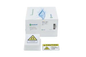
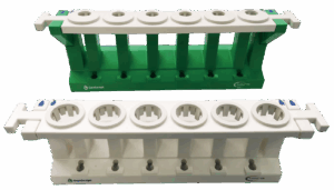
AmMag™ Block
$266.51 Add to cart View Product DetailsGenScript’s AmMag Block
enables protein and antibody purification from cultures up to 1 L in volume
using the AmMag SA. Magnetic beads are directly incubated in cultures or
lysates to bind the target proteins or antibodies. Once binding is complete,
the AmMag Block is used to collect the beads at the bottom of the culture
vessel, and the supernatant is discarded. The magnetic beads thus collected are
re-suspended in a smaller volume and transferred into 50 ml tubes, to be washed
and eluted automatically using the AmMag SA purification system, or manually
using the AmMag MR magnetic racks. -
AmMag™ MR magnetic rack
$1,242.86 Add to cart View Product DetailsGenScript AmMagTM MR magnetic separation rack enables mid-scale separation of magnetic beads. The rack is designed to separate GenScript’s paramagnetic beads from solutions contained in 15 mL or 50 mL tubes.
-
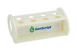
AmMag™ MR-mini magnetic rack
$151.80 Add to cart View Product DetailsGenScript AmMagTM MR-mini magnetic separation rack enables small-scale separation of magnetic beads. The rack is designed to separate GenScript’s paramagnetic beads from solutions contained in 1.5 mL and 2 mL micro-centrifuge tubes. The magnetic field has been optimized for separation of tubes < 2 mL in volume.
-
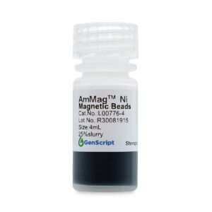
AmMag™ Ni Magnetic beads
$439.88 Add to cart View Product DetailsGenScript’s AmMag™ Ni magnetic beads are designed for purification and screening of histidine-tagged (his-tagged) proteins. The beads are highly EDTA and DTT resistant (Table2), enabling them to bind proteins in the presence of chelators. The beads are also optimized for easy cleanup and regeneration, for reuse with multiple samples. Table 1 describes the basic properties of the AmMag™ Ni magnetic beads.
-

AmMag™ Ni Magnetic beads
$3,415.50 Add to cart View Product DetailsGenScript’s AmMag™ Ni magnetic beads are designed for purification and screening of histidine-tagged (his-tagged) proteins. The beads are highly EDTA and DTT resistant (Table2), enabling them to bind proteins in the presence of chelators. The beads are also optimized for easy cleanup and regeneration, for reuse with multiple samples. Table 1 describes the basic properties of the AmMag™ Ni magnetic beads.
-
AmMag™ Ni Magnetic beads
$25,616.25 Add to cart View Product DetailsGenScript’s AmMag™ Ni magnetic beads are designed for purification and screening of histidine-tagged (his-tagged) proteins. The beads are highly EDTA and DTT resistant (Table2), enabling them to bind proteins in the presence of chelators. The beads are also optimized for easy cleanup and regeneration, for reuse with multiple samples. Table 1 describes the basic properties of the AmMag™ Ni magnetic beads.
-

AmMag™ Protein A Magnetic Beads
$2,285.63 Add to cart View Product DetailsGenScript AmMagᵀᴹ Protein A Magnetic Beads are developed and optimized for antibody purification for high throughput and high volume applications.
The sample containing antibody is added to the AmMagᵀᴹ Protein A Magnetic Beads and incubated on a shaker overnight or 2 hours (depending on the concentration of samples) for the antibody to bind to the beads. The antibody bound to the beads can be eluted off by using acidic elution facilitated by a magnetic separation device. Magnetic separation eliminates the need for centrifugation, minimizes the loss of sample and makes the process more userfriendly.
GenScript AmMagᵀᴹ Protein A Magnetic Beads are super paramagnetic beads with diameter of 45-100μm, covalently coated with alkaline tolerant protein A. The beads are supplied as 25% slurry in 20% ethanol. The AmMagᵀᴹ protein A Magnetic Beads have a binding capacity of 40mghuman IgG per 1 ml settled beads.They can be reused for up to 40 cycles. A total of 20% reduction in binding capacityis observed after 40 cycles.
Protein A, a bacterial cell wall protein isolated from Staphylococcus aureus, binds to mammalian IgG’s, mainly through the Fc regions.The alkaline tolerant protein A not only keeps the specific binding capacity to Fc region of immunoglobulin molecules, but also tolerates alkaline conditions and can withstand rigorous cleaning procedures.
-
AmMag™ Protein A Magnetic Beads
$528.71 Add to cart View Product DetailsGenScript AmMagᵀᴹ Protein A Magnetic Beads are developed and optimized for antibody purification for high throughput and high volume applications.
The sample containing antibody is added to the AmMagᵀᴹ Protein A Magnetic Beads and incubated on a shaker overnight or 2 hours (depending on the concentration of samples) for the antibody to bind to the beads. The antibody bound to the beads can be eluted off by using acidic elution facilitated by a magnetic separation device. Magnetic separation eliminates the need for centrifugation, minimizes the loss of sample and makes the process more userfriendly.
GenScript AmMagᵀᴹ Protein A Magnetic Beads are super paramagnetic beads with diameter of 45-100μm, covalently coated with alkaline tolerant protein A. The beads are supplied as 25% slurry in 20% ethanol. The AmMagᵀᴹ protein A Magnetic Beads have a binding capacity of 40mghuman IgG per 1 ml settled beads.They can be reused for up to 40 cycles. A total of 20% reduction in binding capacityis observed after 40 cycles.
Protein A, a bacterial cell wall protein isolated from Staphylococcus aureus, binds to mammalian IgG’s, mainly through the Fc regions.The alkaline tolerant protein A not only keeps the specific binding capacity to Fc region of immunoglobulin molecules, but also tolerates alkaline conditions and can withstand rigorous cleaning procedures.
-
AmMag™ Protein A Magnetic Beads
$7,719.38 Add to cart View Product DetailsGenScript AmMagᵀᴹ Protein A Magnetic Beads are developed and optimized for antibody purification for high throughput and high volume applications.
-
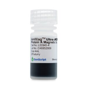
AmMag™ Ultra AT protein A magbeads
$2,285.63 Add to cart View Product DetailsIntended Use
AmMag™ Ultra AT protein A magbeads
are developed and optimized
for antibody purification for high throughput and high volume applications.
Principle
The sample containing
antibody is added to the AmMag™ Ultra AT protein A
magbeads and incubated on a rotor overnight or 2 hours (depending on the
concentration of samples) for the antibody to bind to the beads. The antibody
bound to the beads can be eluted off by using acidic elution facilitated by a
magnetic separation device. Magnetic separation eliminates the need for
centrifugation, minimizes the loss of sample and makes the process more user
friendly.
Material Supplied
GenScript AmMag™ Ultra AT protein A magbeads are super paramagnetic
beads with diameter of 45-100 μm, covalently coated with Ultra alkaline
tolerant protein A, After treatment with 1M NaOH for 24h, the load
decreased by no more than 10%. The beads are supplied as 25% slurry in 20% ethanol.
The AmMag™ Ultra AT protein A
magbeads have a binding capacity of 40mg human IgG per 1 ml settled beads. They
can be reused for up to 100 cycles. After 100 cycles, there was almost no decrease in
binding load.
Protein A, a bacterial cell
wall protein isolated from Staphylococcus aureus, binds to mammalian IgG’s,
mainly through the Fc regions. The alkaline tolerant protein A not only keeps
the specific binding capacity to Fc region of immunoglobulin molecules, but
also tolerates alkaline conditions and can withstand rigorous cleaning
procedures. -
AmMag™ Ultra AT protein A magbeads
$16,818.75 Add to cart View Product DetailsIntended Use
AmMag™ Ultra AT protein A magbeads
are developed and optimized
for antibody purification for high throughput and high volume applications.
Principle
The sample containing
antibody is added to the AmMag™ Ultra AT protein A
magbeads and incubated on a rotor overnight or 2 hours (depending on the
concentration of samples) for the antibody to bind to the beads. The antibody
bound to the beads can be eluted off by using acidic elution facilitated by a
magnetic separation device. Magnetic separation eliminates the need for
centrifugation, minimizes the loss of sample and makes the process more user
friendly.
Material Supplied
GenScript AmMag™ Ultra AT protein A magbeads are super paramagnetic
beads with diameter of 45-100 μm, covalently coated with Ultra alkaline
tolerant protein A, After treatment with 1M NaOH for 24h, the load
decreased by no more than 10%. The beads are supplied as 25% slurry in 20% ethanol.
The AmMag™ Ultra AT protein A
magbeads have a binding capacity of 40mg human IgG per 1 ml settled beads. They
can be reused for up to 100 cycles. After 100 cycles, there was almost no decrease in
binding load.
Protein A, a bacterial cell
wall protein isolated from Staphylococcus aureus, binds to mammalian IgG’s,
mainly through the Fc regions. The alkaline tolerant protein A not only keeps
the specific binding capacity to Fc region of immunoglobulin molecules, but
also tolerates alkaline conditions and can withstand rigorous cleaning
procedures. -
AmMag™ Ultra AT protein A magbeads
$528.71 Add to cart View Product DetailsIntended Use
AmMag™ Ultra AT protein A magbeads
are developed and optimized
for antibody purification for high throughput and high volume applications.
Principle
The sample containing
antibody is added to the AmMag™ Ultra AT protein A
magbeads and incubated on a rotor overnight or 2 hours (depending on the
concentration of samples) for the antibody to bind to the beads. The antibody
bound to the beads can be eluted off by using acidic elution facilitated by a
magnetic separation device. Magnetic separation eliminates the need for
centrifugation, minimizes the loss of sample and makes the process more user
friendly.
Material Supplied
GenScript AmMag™ Ultra AT protein A magbeads are super paramagnetic
beads with diameter of 45-100 μm, covalently coated with Ultra alkaline
tolerant protein A, After treatment with 1M NaOH for 24h, the load
decreased by no more than 10%. The beads are supplied as 25% slurry in 20% ethanol.
The AmMag™ Ultra AT protein A
magbeads have a binding capacity of 40mg human IgG per 1 ml settled beads. They
can be reused for up to 100 cycles. After 100 cycles, there was almost no decrease in
binding load.
Protein A, a bacterial cell
wall protein isolated from Staphylococcus aureus, binds to mammalian IgG’s,
mainly through the Fc regions. The alkaline tolerant protein A not only keeps
the specific binding capacity to Fc region of immunoglobulin molecules, but
also tolerates alkaline conditions and can withstand rigorous cleaning
procedures. -
AmMag™ Ultra AT protein A magbeads
$7,719.38 Add to cart View Product DetailsIntended Use
AmMag™ Ultra AT protein A magbeads
are developed and optimized
for antibody purification for high throughput and high volume applications.
Principle
The sample containing
antibody is added to the AmMag™ Ultra AT protein A
magbeads and incubated on a rotor overnight or 2 hours (depending on the
concentration of samples) for the antibody to bind to the beads. The antibody
bound to the beads can be eluted off by using acidic elution facilitated by a
magnetic separation device. Magnetic separation eliminates the need for
centrifugation, minimizes the loss of sample and makes the process more user
friendly.
Material Supplied
GenScript AmMag™ Ultra AT protein A magbeads are super paramagnetic
beads with diameter of 45-100 μm, covalently coated with Ultra alkaline
tolerant protein A, After treatment with 1M NaOH for 24h, the load
decreased by no more than 10%. The beads are supplied as 25% slurry in 20% ethanol.
The AmMag™ Ultra AT protein A
magbeads have a binding capacity of 40mg human IgG per 1 ml settled beads. They
can be reused for up to 100 cycles. After 100 cycles, there was almost no decrease in
binding load.
Protein A, a bacterial cell
wall protein isolated from Staphylococcus aureus, binds to mammalian IgG’s,
mainly through the Fc regions. The alkaline tolerant protein A not only keeps
the specific binding capacity to Fc region of immunoglobulin molecules, but
also tolerates alkaline conditions and can withstand rigorous cleaning
procedures. -
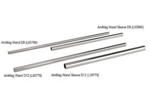
AmMag™ Wand D12
$266.51 Add to cart View Product DetailsGenScript’s AmMag Wand enables protein and antibody purification from cultures up to 2 L in volume using the AmMag SA. Magnetic beads are directly incubated in cultures or lysates to bind target proteins and antibodies. Once binding is complete, the AmMag Wand is used to collect the beads and transfer them into 50 ml tubes, to be washed and eluted automatically using the AmMag SA purification system, or manually using the AmMag MR magnetic racks.
-
AmMag™ Wand D9
$266.51 Add to cart View Product DetailsGenScript’s AmMag Wand enables protein and antibody purification from cultures up to 2 L in volume using the AmMag SA. Magnetic beads are directly incubated in cultures or lysates to bind target proteins and antibodies. Once binding is complete, the AmMag Wand is used to collect the beads and transfer them into 50 ml tubes, to be washed and eluted automatically using the AmMag SA purification system, or manually using the AmMag MR magnetic racks.
-
AmMag™ Wand Sleeve D12
$51.75 Add to cart View Product DetailsGenScript’s AmMag Wand enables protein and antibody purification from cultures up to 2 L in volume using the AmMag SA. Magnetic beads are directly incubated in cultures or lysates to bind target proteins and antibodies. Once binding is complete, the AmMag Wand is used to collect the beads and transfer them into 50 ml tubes, to be washed and eluted automatically using the AmMag SA purification system, or manually using the AmMag MR magnetic racks.
-
AmMag™ Wand Sleeve D9
$47.44 Add to cart View Product DetailsGenScript’s AmMag Wand
enables protein and antibody purification from cultures up to 2 L in volume
using the AmMag SA. Magnetic beads are directly incubated in cultures or
lysates to bind target proteins and antibodies. Once binding is complete, the
AmMag Wand is used to collect the beads and transfer them into 50 ml tubes, to
be washed and eluted automatically using the AmMag SA purification system, or
manually using the AmMag MR magnetic racks. -
Amphiregulin, Human
$43.13 Add to cart View Product DetailsAmphiregulin is a member of the EGF family of cytokines, which comprises at least ten proteins including EGF, TGF-α, HB-EGF, Epiregulin, Tomoregulin, Neuregulins and the various heregulins. Through the EGF/TGF-α receptor, it stimulates growth of keratinocytes, epithelial cells and some fibroblasts. Amphiregulin also inhibits the growth of certain carcinoma cell lines. Synthesized as a transmembrane protein, Amphiregulin’s extracellular domain is proteolytically processed to release the mature protein.
-
Amphiregulin, Human
$86.25 Add to cart View Product DetailsAmphiregulin is a member of the EGF family of cytokines, which comprises at least ten proteins including EGF, TGF-α, HB-EGF, Epiregulin, Tomoregulin, Neuregulins and the various heregulins. Through the EGF/TGF-α receptor, it stimulates growth of keratinocytes, epithelial cells and some fibroblasts. Amphiregulin also inhibits the growth of certain carcinoma cell lines. Synthesized as a transmembrane protein, Amphiregulin’s extracellular domain is proteolytically processed to release the mature protein.
-
Annexin V, His, Human
$63.83 Add to cart View Product DetailsANXA5, also known as annexin V, is a member of the annexin family of calcium-dependent phospholipid binding proteins which are involved in membrane-related activity along exocytotic and endocytotic pathways. ANXA5 is a phospholipase A2 and protein kinase C inhibitory protein with calcium channel properties. It functions in signal transduction, inflammation, cell growth and differentiation. ANXA5 is an anticoagulant protein that acts as an indirect inhibitor of the thromboplastin-specific complex, which is involved in the blood coagulation cascade. There are at least ten different annexins in the mammalian species. Annexins do not contain signal peptides, yet some annexins (A1, A2 and A5) appear to be secreted in a physiologically regulated fashion.The binding of labeled ANXA5 to phosphatidylserine is commonly used as a marker of apoptosis.
-
Annexin V, His, Human
$142.31 Add to cart View Product DetailsANXA5, also known as annexin V, is a member of the annexin family of calcium-dependent phospholipid binding proteins which are involved in membrane-related activity along exocytotic and endocytotic pathways. ANXA5 is a phospholipase A2 and protein kinase C inhibitory protein with calcium channel properties. It functions in signal transduction, inflammation, cell growth and differentiation. ANXA5 is an anticoagulant protein that acts as an indirect inhibitor of the thromboplastin-specific complex, which is involved in the blood coagulation cascade. There are at least ten different annexins in the mammalian species. Annexins do not contain signal peptides, yet some annexins (A1, A2 and A5) appear to be secreted in a physiologically regulated fashion.The binding of labeled ANXA5 to phosphatidylserine is commonly used as a marker of apoptosis.
-
![Anti-AAV2 (intact particle) Antibody (2C4) [Biotin], mAb, Mouse](https://advatechgroup.com/wp-content/uploads/2025/06/antibody01-300x200.jpg)
Anti-AAV2 (intact particle) Antibody (2C4) [Biotin], mAb, Mouse
$207.00 Add to cart View Product DetailsThis antibody recognizes a conformational epitope of assembled AAV2 capsids. It can’t recognize denatured capsid proteins and unassembled capsid proteins.
-
Anti-AAV2 (intact particle) Antibody (2C4), mAb, Mouse
$207.00 Add to cart View Product DetailsThis antibody recognizes a conformational epitope of assembled AAV2 capsids. It can’t recognize denatured capsid proteins and unassembled capsid proteins.
-
Anti-Abatacept Antibody (CT.E8), mAb, Mouse
$124.20 Add to cart View Product DetailsThis product is specific for Abatacept
-
Anti-Abatacept Antibody (CT.F3), mAb, Mouse
$124.20 Add to cart View Product DetailsThis product is specific for Abatacept
-
Anti-Adalimumab Antibody (15C7), mAb, Mouse
$127.65 Add to cart View Product DetailsThe product is specific for Adalimumab. The antibody is recommended as a capture antibody in a pharmacokinetic (PK) bridging assay with detection antibody GenScript, A01956-40, Anti- Adalimumab Antibody (3C2) [Biotin], mAb, Mouse.
-
Anti-Adalimumab Antibody (3C2) [Biotin], mAb, Mouse
$127.65 Add to cart View Product DetailsThe product is specific for Adalimumab. The antibody is recommended as a detection antibody in a pharmacokinetic (PK) bridging assay with capture antibody GenScript, A01954-40, Anti- Adalimumab Antibody (15C7), mAb, Mouse.
-
Anti-Adalimumab Antibody (3C2), mAb, Mouse
$127.65 Add to cart View Product DetailsThe product is specific for Adalimumab. The antibody is recommended as a detection antibody in a pharmacokinetic (PK) bridging assay with capture antibody GenScript, A01954-40, Anti- Adalimumab Antibody (15C7), mAb, Mouse.
-
Anti-Adalimumab Antibody, pAb, Rabbit
$181.99 Add to cart View Product DetailsThe product is specific for Adalimumab. This antibody serves as an excellent positive control for Adalimumab The product is specific for Adalimumab. This antibody serves as an excellent positive control for Adalimumab immunogenicity (ADA) assays.
-
Anti-Atezolizumab Antibody (10G9), mAb, Mouse
$127.65 Add to cart View Product DetailsThe product is specific for Atezolizumab. The antibody is recommended as a capture antibody in a pharmacokinetic (PK) bridging assay with detection antibody GenScript, A01950-40, Anti-Atezolizumab Antibody (6B12) [Biotin], mAb, Mouse.
-
Anti-Atezolizumab Antibody (6B12) [Biotin], mAb, Mouse
$127.65 Add to cart View Product DetailsThe product is specific for Atezolizumab. The antibody is recommended as a detection antibody in a pharmacokinetic (PK) bridging assay with capture antibody GenScript, A01948-40, Anti-Atezolizumab Antibody (10G9), mAb, Mouse.
-
Anti-Atezolizumab Antibody (6B12), mAb, Mouse
$127.65 Add to cart View Product DetailsThe product is specific for Atezolizumab. The antibody is recommended as a detection antibody in a pharmacokinetic (PK) bridging assay with capture antibody GenScript, A01948-40, Anti-Atezolizumab Antibody (10G9), mAb, Mouse.
-
Anti-Avelumab Antibody (12G9), mAb, Mouse
$127.65 Add to cart View Product DetailsThis product is specific for Avelumab
-
Anti-Avelumab Antibody (12H7), mAb, Mouse
$127.65 Add to cart View Product DetailsThis product is specific for Avelumab
-
Anti-Bevacizumab Antibody (11C1), mAb, Mouse
$127.65 Add to cart View Product DetailsThe product is specific for Bevacizumab. The antibody is recommended as a capture antibody in a pharmacokinetic (PK) bridging assay with detection antibody MonoRab™ Anti-Bevacizumab Antibody (46E3), mAb, Rabbit (GenScript, A01962) or MonoRab™ Anti-Bevacizumab Antibody (46E3)[Biotin], mAb, Rabbit (GenScript, A01896) .
-
Anti-Bevacizumab Antibody (4H1), mAb, Mouse
$127.65 Add to cart View Product DetailsThe product is specific for Bevacizumab. The antibody is recommended as a capture antibody in a pharmacokinetic (PK) bridging assay with detection antibody GenScript, A01976-40, Anti-Bevacizumab Antibody (6C3), mAb, Mouse.