GenScript Biotech
Showing 2501–2550 of 2554 results
-

TSH (CT1), mAb, Mouse
$991.88 Add to cart View Product DetailsThyroid stimulating hormone (TSH) is a pituitary hormone that stimulates the thyroid gland to secrete thyroxine (T4) and triiodothyronine (T3). It is a glycoprotein, consisting of the alpha and the beta subunit. TSH serves as a sensitive and specific biomarker for evaluating thyroid function. It is a useful marker for monitoring patients under substitution therapy.
-

TSH (CT1), mAb, Mouse
$8,452.50 Add to cart View Product DetailsThyroid stimulating hormone (TSH) is a pituitary hormone that stimulates the thyroid gland to secrete thyroxine (T4) and triiodothyronine (T3). It is a glycoprotein, consisting of the alpha and the beta subunit. TSH serves as a sensitive and specific biomarker for evaluating thyroid function. It is a useful marker for monitoring patients under substitution therapy.
-

TSHR Antibody (M22HC)
$382.95 Add to cart View Product DetailsThe thyrotropin receptor (TSHR) is the major autoantigen in autoimmune hyperthyroidism. TSHR autoantibodies, acting as TSHR agonists (TRAb), are responsible for Graves’ disease. Measurement of TRAb is important for therapeutic and prognostic purposes.
-

TSHR Antibody (M22HC)
$3,829.50 Add to cart View Product DetailsThe thyrotropin receptor (TSHR) is the major autoantigen in autoimmune hyperthyroidism. TSHR autoantibodies, acting as TSHR agonists (TRAb), are responsible for Graves’ disease. Measurement of TRAb is important for therapeutic and prognostic purposes.
-

TSHR Antibody (M22HC)
$32,583.53 Add to cart View Product DetailsThe thyrotropin receptor (TSHR) is the major autoantigen in autoimmune hyperthyroidism. TSHR autoantibodies, acting as TSHR agonists (TRAb), are responsible for Graves’ disease. Measurement of TRAb is important for therapeutic and prognostic purposes.
-
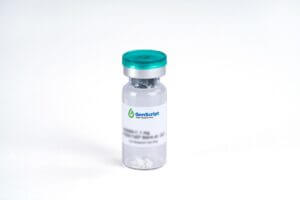
TSLP, Human
$3,820.88 Add to cart View Product DetailsTSLP is a hemopoietic protein that is expressed in the heart, liver and prostate. TSLP overlaps biological activities with IL-7 and binds with the heterodimeric receptor complex consisting of the IL-7R alpha chain (IL-7Rα) and the TSLP-specific chain (TSLPR). Like IL-7, TSLP induces phosphorylation of STAT3 and STAT5, but uses kinases other than the JAKs for activation. TSLP prohibited apoptosis and stimulated growth of the human acute myeloid leukemia (AML)-derived cell line MUTZ3. It induces the release of T cell-attracting chemokines TARC and MDC from monocytes and activates CD11c(+) dendritic cells (DCs). TSLP activated DCs primed naive T cells to produce the proallergic cytokines (IL-4, IL-5, IL-13, TNFα) while down-regulating IL-10 and IFN-γ suggesting a role in initiating allergic inflammation.
-
TSLP, Human
$163.88 Add to cart View Product DetailsTSLP is a hemopoietic protein that is expressed in the heart, liver and prostate. TSLP overlaps biological activities with IL-7 and binds with the heterodimeric receptor complex consisting of the IL-7R alpha chain (IL-7Rα) and the TSLP-specific chain (TSLPR). Like IL-7, TSLP induces phosphorylation of STAT3 and STAT5, but uses kinases other than the JAKs for activation. TSLP prohibited apoptosis and stimulated growth of the human acute myeloid leukemia (AML)-derived cell line MUTZ3. It induces the release of T cell-attracting chemokines TARC and MDC from monocytes and activates CD11c(+) dendritic cells (DCs). TSLP activated DCs primed naive T cells to produce the proallergic cytokines (IL-4, IL-5, IL-13, TNFα) while down-regulating IL-10 and IFN-γ suggesting a role in initiating allergic inflammation.
-

Tube
$62.96 Add to cart View Product DetailsTube for eStain L1, eStain L1C, eStain LG,eBlot L1, and eBlot LG.
-

TWEAK, Human
$1,073.81 Add to cart View Product DetailsTWEAK, short for TNF-related weak inducer of apoptosis, is also known as TNFSF12 and DR3LG. It is a type II transmembrane protein belonging to the TNF superfamily. It is expressed widely in many tissues, such as the heart, skeletal muscle, spleen and peripheral blood. Binding of TWEAK to its receptor TWEAKR induces NF-κB activation, chemokine secretion and apoptosis in certain cell types. TWEAK has also been reported to promote endothelial cell proliferation and migration, thus serving as a regulator of angiogenesis.
-

TWEAK, Human
$63.83 Add to cart View Product DetailsTWEAK, short for TNF-related weak inducer of apoptosis, is also known as TNFSF12 and DR3LG. It is a type II transmembrane protein belonging to the TNF superfamily. It is expressed widely in many tissues, such as the heart, skeletal muscle, spleen and peripheral blood. Binding of TWEAK to its receptor TWEAKR induces NF-κB activation, chemokine secretion and apoptosis in certain cell types. TWEAK has also been reported to promote endothelial cell proliferation and migration, thus serving as a regulator of angiogenesis.
-

TWEAK, Human
$155.25 Add to cart View Product DetailsTWEAK, short for TNF-related weak inducer of apoptosis, is also known as TNFSF12 and DR3LG. It is a type II transmembrane protein belonging to the TNF superfamily. It is expressed widely in many tissues, such as the heart, skeletal muscle, spleen and peripheral blood. Binding of TWEAK to its receptor TWEAKR induces NF-κB activation, chemokine secretion and apoptosis in certain cell types. TWEAK has also been reported to promote endothelial cell proliferation and migration, thus serving as a regulator of angiogenesis.
-

Ty1-tag Antibody, pAb, Rabbit
$87.98 Add to cart View Product DetailsThis Antibody recognizes both Ty1 tagged fusion proteins.
-
![V5-tag Antibody [HRP], pAb, Rabbit](https://advatechgroup.com/wp-content/uploads/2025/06/antibody01-300x200.jpg.pagespeed.ce.lJjhncTAx7.jpg)
V5-tag Antibody [HRP], pAb, Rabbit
$87.98 Add to cart View Product DetailsGenScript Rabbit Anti-V5-tag [HRP] Polyclonal Antibody specifically recognizes V5 tagged fusion proteins.
-

V5-tag Antibody, pAb, Rabbit
$87.98 Add to cart View Product DetailsGenScript Rabbit Anti-V5-tag Polyclonal Antibody specifically recognizes V5 tagged fusion proteins.
-
VEGF-A164, Mouse
$1,470.56 Add to cart View Product DetailsVascular Endothelial Growth Factor (VEGF) was initially purified from media conditioned by normal bovine pituitary folliculo-stellate cells and by a variety of transformed cell lines as a mitogen specific for vascular endothelial cells. It was subsequently found to be identical to an independently discovered vascular permeability factor (VPF), which was previously identified in media conditioned by tumor cell lines based on its ability to increase the permeability of capillary blood vessels. Three mouse cDNA clones, which arise through alternative splicing and which encode mature mouse monomeric VEGF having 120, 164, or 188, amino acids, respectively, have been identified. Two receptor tyrosine kinases (RTKs), Flt-1 and Flk-1 (the mouse homologue of human KDR), both members of the type III subclass of RTKs containing seven immunoglobulin-like repeats in their extracellular domains, have been shown to bind VEGF with high affinity. The roles of the homodimers of KDR, Flt, and the heterodimer of KDR/Flt in VEGF signal transduction remain to be elucidated. In vivo, VEGF has been found to be a potent angiogenesis inducer.
-

VEGF-A164, Mouse
$86.25 Add to cart View Product DetailsVascular Endothelial Growth Factor (VEGF) was initially purified from media conditioned by normal bovine pituitary folliculo-stellate cells and by a variety of transformed cell lines as a mitogen specific for vascular endothelial cells. It was subsequently found to be identical to an independently discovered vascular permeability factor (VPF), which was previously identified in media conditioned by tumor cell lines based on its ability to increase the permeability of capillary blood vessels. Three mouse cDNA clones, which arise through alternative splicing and which encode mature mouse monomeric VEGF having 120, 164, or 188, amino acids, respectively, have been identified. Two receptor tyrosine kinases (RTKs), Flt-1 and Flk-1 (the mouse homologue of human KDR), both members of the type III subclass of RTKs containing seven immunoglobulin-like repeats in their extracellular domains, have been shown to bind VEGF with high affinity. The roles of the homodimers of KDR, Flt, and the heterodimer of KDR/Flt in VEGF signal transduction remain to be elucidated. In vivo, VEGF has been found to be a potent angiogenesis inducer.
-

VEGF-A164, Mouse
$194.06 Add to cart View Product DetailsVascular Endothelial Growth Factor (VEGF) was initially purified from media conditioned by normal bovine pituitary folliculo-stellate cells and by a variety of transformed cell lines as a mitogen specific for vascular endothelial cells. It was subsequently found to be identical to an independently discovered vascular permeability factor (VPF), which was previously identified in media conditioned by tumor cell lines based on its ability to increase the permeability of capillary blood vessels. Three mouse cDNA clones, which arise through alternative splicing and which encode mature mouse monomeric VEGF having 120, 164, or 188, amino acids, respectively, have been identified. Two receptor tyrosine kinases (RTKs), Flt-1 and Flk-1 (the mouse homologue of human KDR), both members of the type III subclass of RTKs containing seven immunoglobulin-like repeats in their extracellular domains, have been shown to bind VEGF with high affinity. The roles of the homodimers of KDR, Flt, and the heterodimer of KDR/Flt in VEGF signal transduction remain to be elucidated. In vivo, VEGF has been found to be a potent angiogenesis inducer.
-

VEGF-C, Human
$1,323.94 Add to cart View Product DetailsVascular endothelial growth factor C (VEGF-C) is a member of the platelet-derived growth factor/vascular endothelial growth factor (PDGF/VEGF) family, is active in angiogenesis, lymphangiogenesis and endothelial cell growth and survival, and can also affect the permeability of blood vessels. VEGF-C is expressed in various tissues, however it is not produced in peripheral blood lymphocytes. It forms cell surface-associated non-covalent disulfide linked homodimers, and can bind and activate both VEGFR-2 (flk1) and VEGFR-3 (flt4) receptors. The structure and function of VEGF-C is similar to those of vascular endothelial growth factor D (VEGF-D).
-

VEGF-C, Human
$63.83 Add to cart View Product DetailsVascular endothelial growth factor C (VEGF-C) is a member of the platelet-derived growth factor/vascular endothelial growth factor (PDGF/VEGF) family, is active in angiogenesis, lymphangiogenesis and endothelial cell growth and survival, and can also affect the permeability of blood vessels. VEGF-C is expressed in various tissues, however it is not produced in peripheral blood lymphocytes. It forms cell surface-associated non-covalent disulfide linked homodimers, and can bind and activate both VEGFR-2 (flk1) and VEGFR-3 (flt4) receptors. The structure and function of VEGF-C is similar to those of vascular endothelial growth factor D (VEGF-D).
-

VEGF-C, Human
$155.25 Add to cart View Product DetailsVascular endothelial growth factor C (VEGF-C) is a member of the platelet-derived growth factor/vascular endothelial growth factor (PDGF/VEGF) family, is active in angiogenesis, lymphangiogenesis and endothelial cell growth and survival, and can also affect the permeability of blood vessels. VEGF-C is expressed in various tissues, however it is not produced in peripheral blood lymphocytes. It forms cell surface-associated non-covalent disulfide linked homodimers, and can bind and activate both VEGFR-2 (flk1) and VEGFR-3 (flt4) receptors. The structure and function of VEGF-C is similar to those of vascular endothelial growth factor D (VEGF-D).
-

VEGF-D, Human
$2,018.25 Add to cart View Product DetailsVascular Endothelial Growth Factor (VEGF)-D, also known as c-Fos-induced growth factor (FIGF), is a member of the PDGF/VEGF growth factor family. It is expressed highly in lung, heart and small intestine, and at lower levels in skeletal muscle, colon and pancreas. It binds to VEGFR-2 and VEGFR-3 receptors and activates downstream signals. VEGF-D is a growth factor active in angiogenesis, lymphangiogenesis and endothelial cell growth. It is involved in many developmental and physiological processes including the formation of venous and lymphatic vascular systems during embryogenesis and the maintenance of differentiated lymphatic endothelium in adults. In tumor pathology, it has been reported to play a role in restructuring of lymphatic channels and regional lymph node metastasis.
-

VEGF-D, Human
$94.88 Add to cart View Product DetailsVascular Endothelial Growth Factor (VEGF)-D, also known as c-Fos-induced growth factor (FIGF), is a member of the PDGF/VEGF growth factor family. It is expressed highly in lung, heart and small intestine, and at lower levels in skeletal muscle, colon and pancreas. It binds to VEGFR-2 and VEGFR-3 receptors and activates downstream signals. VEGF-D is a growth factor active in angiogenesis, lymphangiogenesis and endothelial cell growth. It is involved in many developmental and physiological processes including the formation of venous and lymphatic vascular systems during embryogenesis and the maintenance of differentiated lymphatic endothelium in adults. In tumor pathology, it has been reported to play a role in restructuring of lymphatic channels and regional lymph node metastasis.
-

VEGF-D, Human
$271.69 Add to cart View Product DetailsVascular Endothelial Growth Factor (VEGF)-D, also known as c-Fos-induced growth factor (FIGF), is a member of the PDGF/VEGF growth factor family. It is expressed highly in lung, heart and small intestine, and at lower levels in skeletal muscle, colon and pancreas. It binds to VEGFR-2 and VEGFR-3 receptors and activates downstream signals. VEGF-D is a growth factor active in angiogenesis, lymphangiogenesis and endothelial cell growth. It is involved in many developmental and physiological processes including the formation of venous and lymphatic vascular systems during embryogenesis and the maintenance of differentiated lymphatic endothelium in adults. In tumor pathology, it has been reported to play a role in restructuring of lymphatic channels and regional lymph node metastasis.
-

VEGF-R2 Fc Chimera, Mouse
$1,035.00 Add to cart View Product DetailsVEGF-R2 belongs to a family of proteins called receptor tyrosine kinases. The receptor has three main parts: one part extends out of the cell and binds to VEGF, another spans the cell’s membrane, while the third part is found inside the cell. The current model of VEGF-R2 activation is that VEGF binds to individual VEGF-R2 receptor proteins on the membrane, and brings two of them close enough to form a complex called a dimer. The receptor dimer is activated and initiates signaling within the cell. VEGF-R2 is a receptor tyrosine kinase (RTK) which transduces biochemical signals via lateral dimerization in the plasma membrane. Like most RTKs, VEGF-R2 is composed of an extracellular (EC) domain, a transmembrane (TM) domain, and an intracellular (IC) domain consisting of a kinase domain and sequences required for downstream signaling. The EC domain consists of seven immunoglobulin homology (Ig) domains, termed D1 (at the N-terminus) to D7 (closest to the membrane). VEGF-R2 binds to, and is activated by the ligands VEGF-A, VEGF-E, and a number of processed forms of VEGF-C and VEGF-D. Ligand binding to VEGF-R2 is mediated by Ig-domains 2 and 3 and the linker between D2 and D3.
-

VEGF-R2 Fc Chimera, Mouse
$172.50 Add to cart View Product DetailsVEGF-R2 belongs to a family of proteins called receptor tyrosine kinases. The receptor has three main parts: one part extends out of the cell and binds to VEGF, another spans the cell’s membrane, while the third part is found inside the cell. The current model of VEGF-R2 activation is that VEGF binds to individual VEGF-R2 receptor proteins on the membrane, and brings two of them close enough to form a complex called a dimer. The receptor dimer is activated and initiates signaling within the cell. VEGF-R2 is a receptor tyrosine kinase (RTK) which transduces biochemical signals via lateral dimerization in the plasma membrane. Like most RTKs, VEGF-R2 is composed of an extracellular (EC) domain, a transmembrane (TM) domain, and an intracellular (IC) domain consisting of a kinase domain and sequences required for downstream signaling. The EC domain consists of seven immunoglobulin homology (Ig) domains, termed D1 (at the N-terminus) to D7 (closest to the membrane). VEGF-R2 binds to, and is activated by the ligands VEGF-A, VEGF-E, and a number of processed forms of VEGF-C and VEGF-D. Ligand binding to VEGF-R2 is mediated by Ig-domains 2 and 3 and the linker between D2 and D3.
-

VEGF-R2 Fc Chimera, Mouse
$103.50 Add to cart View Product DetailsVEGF-R2 belongs to a family of proteins called receptor tyrosine kinases. The receptor has three main parts: one part extends out of the cell and binds to VEGF, another spans the cell’s membrane, while the third part is found inside the cell. The current model of VEGF-R2 activation is that VEGF binds to individual VEGF-R2 receptor proteins on the membrane, and brings two of them close enough to form a complex called a dimer. The receptor dimer is activated and initiates signaling within the cell. VEGF-R2 is a receptor tyrosine kinase (RTK) which transduces biochemical signals via lateral dimerization in the plasma membrane. Like most RTKs, VEGF-R2 is composed of an extracellular (EC) domain, a transmembrane (TM) domain, and an intracellular (IC) domain consisting of a kinase domain and sequences required for downstream signaling. The EC domain consists of seven immunoglobulin homology (Ig) domains, termed D1 (at the N-terminus) to D7 (closest to the membrane). VEGF-R2 binds to, and is activated by the ligands VEGF-A, VEGF-E, and a number of processed forms of VEGF-C and VEGF-D. Ligand binding to VEGF-R2 is mediated by Ig-domains 2 and 3 and the linker between D2 and D3.
-

VEGF120, Mouse
$3,187.80 Add to cart View Product DetailsVEGF was initially purified from media conditioned by normal bovine pituitary folliculo-stellate cells and by a variety of transformed cell lines as a mitogen specific for vascular endothelial cells. It was subsequently found to be identical to an independently discovered vascular permeability factor (VPF), which was previously identified in media conditioned by tumor cell lines based on its ability to increase the permeability of capillary blood vessels. Three mouse cDNA clones, which arise through alternative splicing and which encode mature mouse monomeric VEGF having 120, 164, or 188, amino acids, respectively, have been identified. Two receptor tyrosine kinases (RTKs), Flt-1 and Flk-1 (the mouse homologue of human KDR), both members of the type III subclass of RTKs containing seven immunoglobulin-like repeats in their extracellular domains, have been shown to bind VEGF with high affinity. The roles of the homodimers of KDR, Flt, and the heterodimer ofKDR/Flt in VEGF signal transduction remain to be elucidated.In vivo, VEGF has been found to be a potent angiogenesis inducer.
-

VEGF120, Mouse
$194.93 Add to cart View Product DetailsVEGF was initially purified from media conditioned by normal bovine pituitary folliculo-stellate cells and by a variety of transformed cell lines as a mitogen specific for vascular endothelial cells. It was subsequently found to be identical to an independently discovered vascular permeability factor (VPF), which was previously identified in media conditioned by tumor cell lines based on its ability to increase the permeability of capillary blood vessels. Three mouse cDNA clones, which arise through alternative splicing and which encode mature mouse monomeric VEGF having 120, 164, or 188, amino acids, respectively, have been identified. Two receptor tyrosine kinases (RTKs), Flt-1 and Flk-1 (the mouse homologue of human KDR), both members of the type III subclass of RTKs containing seven immunoglobulin-like repeats in their extracellular domains, have been shown to bind VEGF with high affinity. The roles of the homodimers of KDR, Flt, and the heterodimer ofKDR/Flt in VEGF signal transduction remain to be elucidated.In vivo, VEGF has been found to be a potent angiogenesis inducer.
-

VEGF121, Human
$86.25 Add to cart View Product DetailsVEGF-A121 is one of five isoforms (121, 145, 165, 189, and 206) of VEGF protein, a cytokine belonging to the Platelet Differentiation Growth Factor (PDGF) family, and existing as a disulfide-linked homodimeric glycoprotein. In contrast to the longer isoforms, VEGF-A121 is more freely diffusible, and cannot bind to heparin. In vivo, VEGF is expressed predominantly in lung, heart, kidney, and adrenal glands, and the expression of VEGF is up-regulated by a number of growth factors, including PDGF, Fibroblast Growth Factor (FGF), Epidermal Growth Factor (EGF), and Tumor Necrosis Factor (TNF). VEGF signals via binding to two tyrosine kinase receptors: the Fms-like tyrosine kinase 1 (Flt-1) and the kinase domain receptor (KDR). VEGF is a specific mitogen and survival factor, contributing to abnormal angiogenesis and cancer development.
-

VEGF121, Human
$271.69 Add to cart View Product DetailsVEGF-A121 is one of five isoforms (121, 145, 165, 189, and 206) of VEGF protein, a cytokine belonging to the Platelet Differentiation Growth Factor (PDGF) family, and existing as a disulfide-linked homodimeric glycoprotein. In contrast to the longer isoforms, VEGF-A121 is more freely diffusible, and cannot bind to heparin. In vivo, VEGF is expressed predominantly in lung, heart, kidney, and adrenal glands, and the expression of VEGF is up-regulated by a number of growth factors, including PDGF, Fibroblast Growth Factor (FGF), Epidermal Growth Factor (EGF), and Tumor Necrosis Factor (TNF). VEGF signals via binding to two tyrosine kinase receptors: the Fms-like tyrosine kinase 1 (Flt-1) and the kinase domain receptor (KDR). VEGF is a specific mitogen and survival factor, contributing to abnormal angiogenesis and cancer development.
-

VEGF164, Mouse
$155.25 Add to cart View Product DetailsVascular Endothelial Growth Factor (VEGF) was initially purified from media conditioned by normal bovine pituitary folliculo-stellate cells and by a variety of transformed cell lines as a mitogen specific for vascular endothelial cells. It was subsequently found to be identical to an independently discovered vascular permeability factor (VPF), which was previously identified in media conditioned by tumor cell lines based on its ability to increase the permeability of capillary blood vessels. Three mouse cDNA clones, which arise through alternative splicing and which encode mature mouse monomeric VEGF having 120, 164, or 188, amino acids, respectively, have been identified. Two receptor tyrosine kinases (RTKs), Flt-1 and Flk-1 (the mouse homologue of human KDR), both members of the type III subclass of RTKs containing seven immunoglobulin-like repeats in their extracellular domains, have been shown to bind VEGF with high affinity. The roles of the homodimers of KDR, Flt, and the heterodimer of KDR/Flt in VEGF signal transduction remain to be elucidated. In vivo, VEGF has been found to be a potent angiogenesis inducer.
-

VEGF164, Mouse (P. pastoris-expressed)
$1,470.56 Add to cart View Product DetailsVascular Endothelial Growth Factor (VEGF) was initially purified from media conditioned by normal bovine pituitary folliculo-stellate cells and by a variety of transformed cell lines as a mitogen specific for vascular endothelial cells. It was subsequently found to be identical to an independently discovered vascular permeability factor (VPF), which was previously identified in media conditioned by tumor cell lines based on its ability to increase the permeability of capillary blood vessels. Three mouse cDNA clones, which arise through alternative splicing and which encode mature mouse monomeric VEGF having 120, 164, or 188, amino acids, respectively, have been identified. Two receptor tyrosine kinases (RTKs), Flt-1 and Flk-1 (the mouse homologue of human KDR), both members of the type III subclass of RTKs containing seven immunoglobulin-like repeats in their extracellular domains, have been shown to bind VEGF with high affinity. The roles of the homodimers of KDR, Flt, and the heterodimer of KDR/Flt in VEGF signal transduction remain to be elucidated. In vivo, VEGF has been found to be a potent angiogenesis inducer.
-

VEGF164, Rat (CHO-expressed)
$1,470.56 Add to cart View Product DetailsVascular Endothelial Growth Factor A164 (VEGF-A164), a member of the cysteine knot growth factor, is one of major isoforms of VEGF-As. VEGF-As are endothelial cell-specific mitogens with angiogenic and vascular permeability-inducing properties. During maturation, rat VEGF-A is alternatively spliced to generate rVEGF-A120, rVEGF-A164 and rVEGF-A188 which correspond to hVEGF-A121, hVEGF-A165 and hVEGF-A189 in human, respectively (the numbers designate the amino acid residues). The active form of rVEGF-A164 is either a homodimeric or heterodimeric polypeptides which bind to the transmembrane tyrosine kinases receptors FLT1, FLK1 or KDR or to the non-tyrosine kinase neuropilin receptors NRP1/2.
-

VEGF164, Rat (CHO-expressed)
$86.25 Add to cart View Product DetailsVascular Endothelial Growth Factor A164 (VEGF-A164), a member of the cysteine knot growth factor, is one of major isoforms of VEGF-As. VEGF-As are endothelial cell-specific mitogens with angiogenic and vascular permeability-inducing properties. During maturation, rat VEGF-A is alternatively spliced to generate rVEGF-A120, rVEGF-A164 and rVEGF-A188 which correspond to hVEGF-A121, hVEGF-A165 and hVEGF-A189 in human, respectively (the numbers designate the amino acid residues). The active form of rVEGF-A164 is either a homodimeric or heterodimeric polypeptides which bind to the transmembrane tyrosine kinases receptors FLT1, FLK1 or KDR or to the non-tyrosine kinase neuropilin receptors NRP1/2.
-

VEGF164, Rat (CHO-expressed)
$194.06 Add to cart View Product DetailsVascular Endothelial Growth Factor A164 (VEGF-A164), a member of the cysteine knot growth factor, is one of major isoforms of VEGF-As. VEGF-As are endothelial cell-specific mitogens with angiogenic and vascular permeability-inducing properties. During maturation, rat VEGF-A is alternatively spliced to generate rVEGF-A120, rVEGF-A164 and rVEGF-A188 which correspond to hVEGF-A121, hVEGF-A165 and hVEGF-A189 in human, respectively (the numbers designate the amino acid residues). The active form of rVEGF-A164 is either a homodimeric or heterodimeric polypeptides which bind to the transmembrane tyrosine kinases receptors FLT1, FLK1 or KDR or to the non-tyrosine kinase neuropilin receptors NRP1/2.
-

VEGF165, Human
$1,470.56 Add to cart View Product DetailsVascular Endothelial Growth Factor (VEGF) is a potent growth and angiogenic cytokine. It stimulates proliferation and survival of endothelial cells, and promotes angiogenesis and vascular permeability. Expressed in vascularized tissues, Vascular Endothelial Growth Factor (VEGF) plays a prominent role in normal and pathological angiogenesis. Substantial evidence implicates Vascular Endothelial Growth Factor (VEGF) in the induction of tumor metastasis and intra-ocular neovascular syndromes. Vascular Endothelial Growth Factor (VEGF) signals through the three receptors; fms-like tyrosine kinase (flt-1), KDR gene product (the murine homolog of KDR is the flk-1 gene product) and the flt4 gene product.
-

VEGF165, Human
$86.25 Add to cart View Product DetailsVascular Endothelial Growth Factor (VEGF) is a potent growth and angiogenic cytokine. It stimulates proliferation and survival of endothelial cells, and promotes angiogenesis and vascular permeability. Expressed in vascularized tissues, Vascular Endothelial Growth Factor (VEGF) plays a prominent role in normal and pathological angiogenesis. Substantial evidence implicates Vascular Endothelial Growth Factor (VEGF) in the induction of tumor metastasis and intra-ocular neovascular syndromes. Vascular Endothelial Growth Factor (VEGF) signals through the three receptors; fms-like tyrosine kinase (flt-1), KDR gene product (the murine homolog of KDR is the flk-1 gene product) and the flt4 gene product.
-

VEGF165, Human
$392.44 Add to cart View Product DetailsVascular Endothelial Growth Factor (VEGF) is a potent growth and angiogenic cytokine. It stimulates proliferation and survival of endothelial cells, and promotes angiogenesis and vascular permeability. Expressed in vascularized tissues, Vascular Endothelial Growth Factor (VEGF) plays a prominent role in normal and pathological angiogenesis. Substantial evidence implicates Vascular Endothelial Growth Factor (VEGF) in the induction of tumor metastasis and intra-ocular neovascular syndromes. Vascular Endothelial Growth Factor (VEGF) signals through the three receptors; fms-like tyrosine kinase (flt-1), KDR gene product (the murine homolog of KDR is the flk-1 gene product) and the flt4 gene product.
-

VEGF165, Human
$271.69 Add to cart View Product DetailsVascular Endothelial Growth Factor (VEGF) is a potent growth and angiogenic cytokine. It stimulates proliferation and survival of endothelial cells, and promotes angiogenesis and vascular permeability. Expressed in vascularized tissues, Vascular Endothelial Growth Factor (VEGF) plays a prominent role in normal and pathological angiogenesis. Substantial evidence implicates Vascular Endothelial Growth Factor (VEGF) in the induction of tumor metastasis and intra-ocular neovascular syndromes. Vascular Endothelial Growth Factor (VEGF) signals through the three receptors; fms-like tyrosine kinase (flt-1), KDR gene product (the murine homolog of KDR is the flk-1 gene product) and the flt4 gene product.
-

VEGF165, Human(HEK 293-expressed)
$1,470.56 Add to cart View Product DetailsVascular Endothelial Growth Factor (VEGF) is a potent growth and angiogenic cytokine. It stimulates proliferation and survival of endothelial cells, and promotes angiogenesis and vascular permeability. Expressed in vascularized tissues, Vascular Endothelial Growth Factor (VEGF) plays a prominent role in normal and pathological angiogenesis. Substantial evidence implicates Vascular Endothelial Growth Factor (VEGF) in the induction of tumor metastasis and intra-ocular neovascular syndromes. Vascular Endothelial Growth Factor (VEGF) signals through the three receptors; fms-like tyrosine kinase (flt-1), KDR gene product (the murine homolog of KDR is the flk-1 gene product) and the flt4 gene product.
-

VEGF165, Human(HEK 293-expressed)
$86.25 Add to cart View Product DetailsVascular Endothelial Growth Factor (VEGF) is a potent growth and angiogenic cytokine. It stimulates proliferation and survival of endothelial cells, and promotes angiogenesis and vascular permeability. Expressed in vascularized tissues, Vascular Endothelial Growth Factor (VEGF) plays a prominent role in normal and pathological angiogenesis. Substantial evidence implicates Vascular Endothelial Growth Factor (VEGF) in the induction of tumor metastasis and intra-ocular neovascular syndromes. Vascular Endothelial Growth Factor (VEGF) signals through the three receptors; fms-like tyrosine kinase (flt-1), KDR gene product (the murine homolog of KDR is the flk-1 gene product) and the flt4 gene product.
-

VEGF165, Human(HEK 293-expressed)
$271.69 Add to cart View Product DetailsVascular Endothelial Growth Factor (VEGF) is a potent growth and angiogenic cytokine. It stimulates proliferation and survival of endothelial cells, and promotes angiogenesis and vascular permeability. Expressed in vascularized tissues, Vascular Endothelial Growth Factor (VEGF) plays a prominent role in normal and pathological angiogenesis. Substantial evidence implicates Vascular Endothelial Growth Factor (VEGF) in the induction of tumor metastasis and intra-ocular neovascular syndromes. Vascular Endothelial Growth Factor (VEGF) signals through the three receptors; fms-like tyrosine kinase (flt-1), KDR gene product (the murine homolog of KDR is the flk-1 gene product) and the flt4 gene product.
-

VHH Affinity Ligand ELISA kit
$733.13 Add to cart View Product DetailsVHH Affinity Ligand ELISA Kit could detect the residual levels of various VHH affinity ligands and also be used for the quantitative analysis of VHH drugs.
-

VISTA/B7-H5 Fc Chimera, Human
$1,035.00 Add to cart View Product DetailsV-domain Ig suppressor of T cell activation (VISTA), also known as B7-H5, is a type I transmembrane protein that functions as an immune checkpoint. VISTA belongs to the immunoglobulin superfamily and has one IgV domain. It is primarily expressed in white blood cells and its transcription is partially controlled by p53. VISTA can act as both a ligand and a receptor on T cells to inhibit T cell effector function and maintain peripheral tolerance. VISTA may also promote differentiation of embryonic stem cells by inhibiting BMP4 signaling (By similarity) and may stimulate MMP14-mediated MMP2 activation.
-

VISTA/B7-H5 Fc Chimera, Human
$172.50 Add to cart View Product DetailsV-domain Ig suppressor of T cell activation (VISTA), also known as B7-H5, is a type I transmembrane protein that functions as an immune checkpoint. VISTA belongs to the immunoglobulin superfamily and has one IgV domain. It is primarily expressed in white blood cells and its transcription is partially controlled by p53. VISTA can act as both a ligand and a receptor on T cells to inhibit T cell effector function and maintain peripheral tolerance. VISTA may also promote differentiation of embryonic stem cells by inhibiting BMP4 signaling (By similarity) and may stimulate MMP14-mediated MMP2 activation.
-

VISTA/B7-H5 Fc Chimera, Human
$103.50 Add to cart View Product DetailsV-domain Ig suppressor of T cell activation (VISTA), also known as B7-H5, is a type I transmembrane protein that functions as an immune checkpoint. VISTA belongs to the immunoglobulin superfamily and has one IgV domain. It is primarily expressed in white blood cells and its transcription is partially controlled by p53. VISTA can act as both a ligand and a receptor on T cells to inhibit T cell effector function and maintain peripheral tolerance. VISTA may also promote differentiation of embryonic stem cells by inhibiting BMP4 signaling (By similarity) and may stimulate MMP14-mediated MMP2 activation.
-
VISTA/B7-H5, His, Human
$1,035.00 Add to cart View Product DetailsV-domain Ig suppressor of T cell activation (VISTA), also known as B7-H5, is a type I transmembrane protein that functions as an immune checkpoint. VISTA belongs to the immunoglobulin superfamily and has one IgV domain. It is primarily expressed in white blood cells and its transcription is partially controlled by p53. VISTA can act as both a ligand and a receptor on T cells to inhibit T cell effector function and maintain peripheral tolerance. VISTA may also promote differentiation of embryonic stem cells by inhibiting BMP4 signaling (By similarity) and may stimulate MMP14-mediated MMP2 activation.
-

VISTA/B7-H5, His, Human
$172.50 Add to cart View Product DetailsV-domain Ig suppressor of T cell activation (VISTA), also known as B7-H5, is a type I transmembrane protein that functions as an immune checkpoint. VISTA belongs to the immunoglobulin superfamily and has one IgV domain. It is primarily expressed in white blood cells and its transcription is partially controlled by p53. VISTA can act as both a ligand and a receptor on T cells to inhibit T cell effector function and maintain peripheral tolerance. VISTA may also promote differentiation of embryonic stem cells by inhibiting BMP4 signaling (By similarity) and may stimulate MMP14-mediated MMP2 activation.
-

VSV-G-tag Antibody, pAb, Rabbit
$87.98 Add to cart View Product DetailsThis Antibody recognizes VSV-G tagged fusion proteins.
-

WB Reaction Box
$14.66 Add to cart View Product DetailsWB Reaction Box
