1mg
Showing 3601–3650 of 3902 results
-
PCT (16A1), mAb, Mouse
$131.10 Add to cart View Product DetailsPCT, a 116 amino acid (aa) protein, is comprised of three sections including a 57 aa N-terminal PCT, a 32 aa calcitonin and a 21 aa katacalcin. Calcitonin is a hormone, derived from PCT cleavage. PCT is a good diagnosis marker for bacterial infection. Other diseases such as sepsis, inflammation, surgery, heat shock, burn injuries and cardiogenic shock can also cause an increase of PCT level in blood.
-
PCT (18D2), mAb, Mouse
$131.10 Add to cart View Product DetailsPCT, a 116 amino acid (aa) protein, is comprised of three sections including a 57 aa N-terminal PCT, a 32 aa calcitonin and a 21 aa katacalcin. Calcitonin is a hormone, derived from PCT cleavage. PCT is a good diagnosis marker for bacterial infection. Other diseases such as sepsis, inflammation, surgery, heat shock, burn injuries and cardiogenic shock can also cause an increase of PCT level in blood.
-
PCT (18M3), mAb, Mouse
$131.10 Add to cart View Product DetailsPCT, a 116 amino acid (aa) protein, is comprised of three sections including a 57 aa N-terminal PCT, a 32 aa calcitonin and a 21 aa katacalcin. Calcitonin is a hormone, derived from PCT cleavage. PCT is a good diagnosis marker for bacterial infection. Other diseases such as sepsis, inflammation, surgery, heat shock, burn injuries and cardiogenic shock can also cause an increase of PCT level in blood.
-
PCT (PE21), mAb, Mouse
$131.10 Add to cart View Product DetailsPCT, a 116 amino acid (aa) protein, is comprised of three sections including a 57 aa N-terminal PCT, a 32 aa calcitonin and a 21 aa katacalcin. Calcitonin is a hormone, derived from PCT cleavage. PCT is a good diagnosis marker for bacterial infection. Other diseases such as sepsis, inflammation, surgery, heat shock, burn injuries and cardiogenic shock can also cause an increase of PCT level in blood.
-
PD-1 (2E5), mAb, Mouse
$1,306.69 Add to cart View Product DetailsProgrammed Cell Death Protein 1 (PD-1), is cell surface receptor expressing on T cells and pro-B cells. The binding of PD-1 to its two ligands, PD-L1 and PD-L2, could result in down-regulation of the immune system by inhibiting the T-cell activation process. Thus, PD-1 is an important immune checkpoint and a popular target for therapeutic antibodies against many cancers.
-

PD-1 Fc Chimera, Human
$1,035.00 Add to cart View Product DetailsProgrammed cell death protein 1, also known as PD-1 and CD279 (cluster of differentiation 279) or PDCD1, is a protein that in humans is encoded by the PDCD1 gene. PD-1 is a cell surface receptor that belongs to the immunoglobulin superfamily and is expressed on T cells and pro-B cells.PD-1 binds two ligands, PD-L1 and PD-L2. PD-1 and its ligands play an important role in down regulating the immune system by preventing the activation of T-cells, which in turn reduces autoimmunity and promotes self-tolerance. The inhibitory effect of PD-1 is accomplished through a dual mechanism of promoting apoptosis (programmed cell death) in antigen specific T-cells in lymph nodes while simultaneously reducing apoptosis in regulatory T cells (suppressor T cells)
-

PD-1 Fc Chimera, Mouse
$1,035.00 Add to cart View Product DetailsProgrammed cell death protein 1, also known as PD-1 and CD279 (cluster of differentiation 279) or PDCD1, is a protein that in humans is encoded by the PDCD1 gene. PD-1 is a cell surface receptor that belongs to the immunoglobulin superfamily and is expressed on T cells and pro-B cells.PD-1 binds two ligands, PD-L1 and PD-L2. PD-1 and its ligands play an important role in down regulating the immune system by preventing the activation of T-cells, which in turn reduces autoimmunity and promotes self-tolerance. The inhibitory effect of PD-1 is accomplished through a dual mechanism of promoting apoptosis (programmed cell death) in antigen specific T-cells in lymph nodes while simultaneously reducing apoptosis in regulatory T cells (suppressor T cells).
-
PD-1, His, Human
$1,293.75 Add to cart View Product DetailsProgrammed death (PD-1) is an immunoinhibitory receptor that belongs to the CD28 family and is expressed on T cells, B cells, monocytes, natural killer cells, and many tumor-infiltrating lymphocytes (TILs); PD-1 is a type I membrane protein of 268 amino acids and which structure includes an extracellular IgV domain followed by a transmembrane region and an intracellular tail. The intracellular tail contains two phosphorylation sites located in an immunoreceptor tyrosine-based inhibitory motif and an immunoreceptor tyrosine-based switch motif, which suggests that PD-1 negatively regulates TCR signals. This is consistent with binding of SHP-1 and SHP-2 phosphatases to the cytoplasmic tail of PD-1 upon ligand binding. It has 2 ligands that have been described PD-L1(B7H1) and PD-L2(B7-DC); PD-1 induction on activated T cells occurs in response to PD-L1 or L2 engagement and limits effector T-cell activity in peripheral organs and tissues during inflammation, thus preventing autoimmunity.Recombinant Human PD-1 produced in HEK293 cells is a polypeptide chain containing 149 amino acids with C-terminal 6×His. A fully biologically active molecule, rhPD-1 has a molecular mass of 30-40 kDa analyzed by reducing SDS-PAGE and is obtained by chromatographic techniques at GenScript.
-
PD-L1 Fc Chimera, Human
$1,035.00 Add to cart View Product DetailsProgrammed death-ligand 1 (PD-L1) also known as cluster of differentiation 274 (CD274) or B7 homolog 1 (B7-H1), is a protein that in humans is encoded by the CD274 gene. PD-L1 is a 40 kDa type 1 transmembrane protein that has been speculated to play a major role in suppressing the immune system during particular events such as pregnancy, tissue allografts, autoimmune disease and other disease states such as hepatitis. Normally the immune system reacts to foreign antigens where there is some accumulation in the lymph nodes or spleen which triggers a proliferation of antigen-specific CD8+ T cell. The formation of PD-1 receptor / PD-L1 or B7.1 receptor /PD-L1 ligand complex transmits an inhibitory signal which reduces the proliferation of these CD8+ T cells at the lymph nodes and supplementary to that PD-1 is also able to control the accumulation of foreign antigen specific T cells in the lymph nodes through apoptosis which is further mediated by a lower regulation of the gene Bcl-2. PD-L1 binds to its receptor, PD-1, found on activated T cells, B cells, and myeloid cells, to modulate activation or inhibition. Recombinant Human PD-L1(B7-H1) Fc Chimera produced in CHO cells is a polypeptide chain containing 457 amino acids. A fully biologically active molecule, rh PD‑L1(B7-H1) has a molecular mass of 70-72 kDa analyzed by reducing SDS-PAGE and is obtained by chromatographic techniques at GenScript.
-
PD-L1 Fc Chimera, Mouse
$1,035.00 Add to cart View Product DetailsProgrammed death-ligand 1 (PD-L1) also known as cluster of differentiation 274 (CD274) or B7 homolog 1 (B7-H1), is a protein that in humans is encoded by the CD274 gene. PD-L1 is a 40 kDa type 1 transmembrane protein that has been speculated to play a major role in suppressing the immune system during particular events such as pregnancy, tissue allografts, autoimmune disease and other disease states such as hepatitis. Normally the immune system reacts to foreign antigens where there is some accumulation in the lymph nodes or spleen which triggers a proliferation of antigen-specific CD8+ T cell. The formation of PD-1 receptor / PD-L1 or B7.1 receptor /PD-L1 ligand complex transmits an inhibitory signal which reduces the proliferation of these CD8+ T cells at the lymph nodes and supplementary to that PD-1 is also able to control the accumulation of foreign antigen specific T cells in the lymph nodes through apoptosis which is further mediated by a lower regulation of the gene Bcl-2. PD-L1 binds to its receptor, PD-1, found on activated T cells, B cells, and myeloid cells, to modulate activation or inhibition.
-

PD-L1, His, Human
$1,035.00 Add to cart View Product DetailsProgrammed death-ligand 1 (PD-L1) also known as cluster of differentiation 274 (CD274) or B7 homolog 1 (B7-H1), is a protein that in humans is encoded by the CD274 gene. PD-L1 is a 40 kDa type 1 transmembrane protein that has been speculated to play a major role in suppressing the immune system during particular events such as pregnancy, tissue allografts, autoimmune disease and other disease states such as hepatitis. Normally the immune system reacts to foreign antigens where there is some accumulation in the lymph nodes or spleen which triggers a proliferation of antigen-specific CD8+ T cell. The formation of PD-1 receptor / PD-L1 or B7.1 receptor /PD-L1 ligand complex transmits an inhibitory signal which reduces the proliferation of these CD8+ T cells at the lymph nodes and supplementary to that PD-1 is also able to control the accumulation of foreign antigen specific T cells in the lymph nodes through apoptosis which is further mediated by a lower regulation of the gene Bcl-2. PD-L1 binds to its receptor, PD-1, found on activated T cells, B cells, and myeloid cells, to modulate activation or inhibition. Recombinant Human PD-L1(B7-H1) Fc Chimera produced in CHO cells is a polypeptide chain containing 457 amino acids. A fully biologically active molecule, rh PD‑L1(B7-H1) has a molecular mass of 70-72 kDa analyzed by reducing SDS-PAGE and is obtained by chromatographic techniques at GenScript.
-

PD-L2 Fc Chimera, Human
$1,035.00 Add to cart View Product DetailsPD-L1 and PD-L2 are ligands for PD-1, a costimulatory molecule that plays an inhibitory role in regulating T cell activation in the periphery. PD-L2 also known as PD-L2, B7-DC serves as a negative and a positive regulator of T cell function. The expression and function of PD-L2 are similar to PD-L1. Both PD-L2−PD-1 and PD-L1−PD-1 signals inhibit T cell proliferation by blocking cell cycle progression but not by increasing cell death. PD-L2−PD-1 interactions are able to inhibit TCR-mediated proliferation and cytokine production in the absence of CD28 costimulation. Threshold for T cell activation may be a balance between activating signals, such as those delivered by the engagement of CD28 by B7-1 and B7-2, and inhibitory signals, mediated by engagement of PD-1 by PD-L1 and PD-L2. The structural conservation of B7-like and CD28-like receptors may reflect the distance between T cells and APCs in the immunological synapse. The PD-L−PD-1 pathway may play a key role in the induction and/or maintenance of peripheral tolerance and autoimmune disease. Because PD-L1 and PD-L2 can inhibit effector T cell proliferation and cytokine production, the PD-L−PD-1 pathway may be an attractive therapeutic target. Blocking the PD-1 pathway may enhance anti-tumor immunity, whereas stimulating this pathway may be useful for down-regulating ongoing immune responses in transplant rejection and autoimmune and allergic diseases.
-

PDGF-AA, Mouse
$1,712.06 Add to cart View Product DetailsPlatelet-Derived Growth Factor-AA (PDGF-AA) is one of five dimers (PDGF-AA, AB, BB, CC, and DD) formed by 4 different PDGF subunits. In chemical terms, platelet-derived growth factor is a dimeric glycoprotein composed of two A (-AA) or two B (-BB) chains or a combination of the two (-AB). The dimeric isoforms PDGFAA, AB and BB are differentially expressed in various cell types, and their effects are mediated through two distinct receptors termed α and β. Differences exist in isoform binding to each receptor. Ingeneral, PDGF isoforms are potent mitogens for connective tissue cells including dermal fibroblasts, glial cells, arterial smooth muscle cells and some epithelial andendothelial cells. In addition to its activity as a mitogen, PDGF is chemotactic for fibroblasts, smooth muscle cells, neutrophils and mononuclear cells. PDGF-AA plays a significant role in blood vessel formation (angiogenesis).
-
PDGF-BB, Bovine
$2,518.50 Add to cart View Product DetailsPlatelet-derived growth factor (PDGF) presenting in serum but absent from plasma was first discovered in an animal study by Lynch and co-workers in the late 1980s. It is a disulfide-linked dimer consisting of two peptides-chain A and chain B. PDGF has three subforms: PDGF-AA, PDGF-BB, and PDGF-AB. It is involved in many biological processes, including hyperplasia, embryonic neuron development, chemotaxis, and respiratory tubule epithelial cell development. The function of PDGF is mediated by two receptors (PDGFR-α and PDGFR-β).
-
PDGF-BB, Human
$2,518.50 Add to cart View Product DetailsPlatelet-derived growth factor (PDGF) presenting in serum but absent from plasma was first discovered in animal study by Lynch and co-workers in the late 1980s. It is a disulfide-linked dimer consisting of two peptides-chain A and chain B. PDGF has three subforms: PDGF-AA, PDGF-BB, PDGF-AB. It is involved in a number of biological processes, including hyperplasia, embryonic neuron development, chemotaxis, and respiratory tubule epithelial cell development. The function of PDGF is mediated by two receptors (PDGFR-α and PDGFR-β).
-
PDGF-BB, Human
$2,596.13 Add to cart View Product DetailsPlatelet-Derived Growth Factor-BB (PDGF-BB) is one of five dimers (PDGF-AA, AB, BB, CC, and DD) formed by 4 different PDGF subunits. In vivo, PDGF-BB is mainly produced in heart and placenta, and predominantly expressed by osteoblasts, fibroblasts, smooth muscle cells, and glial cells. An inactive precursor of PDGF-BB is produced in the endoplasmic reticulum and then activated by a proprotein convertase after secretion. PDGF-BB functions in a paracrine manner and promotes organogenesis, human skeletal development, and wound healing. PDGF-BB also promotes angiogenesis, particularly in the presence of Fibroblast Growth Factor basic. Therefore, PDGF-BB and its related pathways are potential pharmacological targets.
-
PDGF-BB, Human (P. pastoris-expressed)
$2,018.25 Add to cart View Product DetailsPlatelet-Derived Growth Factor-BB (PDGF-BB) is one of five dimers (PDGF-AA, AB, BB, CC, and DD) formed by 4 different PDGF subunits. In vivo, PDGF-BB is mainly produced in heart and placenta, and predominantly expressed by osteoblasts, fibroblasts, smooth muscle cells, and glial cells. An inactive precursor of PDGF-BB is produced in the endoplasmic reticulum and then activated by a proprotein convertase after secretion. PDGF-BB functions in a paracrine manner and promotes organogenesis, human skeletal development, and wound healing. PDGF-BB also promotes angiogenesis, particularly in the presence of Fibroblast Growth Factor basic. Therefore, PDGF-BB and its related pathways are potential pharmacological targets.
-
PDGF-BB, Mouse
$1,470.56 Add to cart View Product DetailsPlatelet-Derived Growth Factor-BB (PDGF-BB) is one of five dimers (PDGF-AA, AB, BB, CC, and DD) formed by 4 different PDGF subunits. In vivo, PDGF-BB is mainly produced in heart and placenta, and predominantly expressed by osteoblasts, fibroblasts, smooth muscle cells, and glial cells. An inactive precursor of PDGF-BB is produced in the endoplasmic reticulum and then activated by a proprotein convertase after secretion. PDGF-BB functions in a paracrine manner and promotes organogenesis, human skeletal development, and wound healing. PDGF-BB also promotes angiogenesis, particularly in the presence of Fibroblast Growth Factor basic. Therefore, PDGF-BB and its related pathways are potential pharmacological targets.
-
PDGF-BB, Mouse
$2,518.50 Add to cart View Product DetailsPlatelet-Derived Growth Factor Subunit B (PDGFB) belongs to the PDGF/VEGF growth factor family. Platelet-derived growth factor is a potent mitogen for cells of mesenchymal origin. PDGFB can exist either as a homodimer (PDGF-BB) or as a heterodimer with the platelet-derived growth factor alpha polypeptide (PDGF-AB), where the dimers are connected by disulfide bonds. As growth factor, it plays an essential role in the regulation of embryonic development, cell proliferation, cell migration, survival and chemotaxis. It is required for normal proliferation and recruitment of pericytes and vascular smooth muscle cells in the central nervous system, skin, lung, heart and placenta. PDGFB also plays an important role in wound healing.
-
PDGF-BB, Rat
$1,470.56 Add to cart View Product DetailsPlatelet-Derived Growth Factor-BB (PDGF-BB) is one of five dimers (PDGF-AA, AB, BB, CC, and DD) formed by 4 different PDGF subunits. In vivo, PDGF-BB is mainly produced in heart and placenta, and predominantly expressed by osteoblasts, fibroblasts, smooth muscle cells, and glial cells. An inactive precursor of PDGF-BB is produced in the endoplasmic reticulum and then activated by a proprotein convertase after secretion. PDGF-BB functions in a paracrine manner and promotes organogenesis, human skeletal development, and wound healing. PDGF-BB also promotes angiogenesis, particularly in the presence of Fibroblast Growth Factor basic. Therefore, PDGF-BB and its related pathways are potential pharmacological targets.
-
PEDF, Human
$2,596.13 Add to cart View Product DetailsPEDF is a noninhibitory serpin with neurotrophic, anti-angiogenic, and anti-tumorigenic properties. It is a 50 kDa glycoprotein produced and secreted in many tissues throughout the body. A major component of the anti-angiogenic action of PEDF is the induction of apoptosis in proliferating endothelial cells. In addition, PEDF is able to inhibit the activity of angiogenic factors such as VEGF and FGF-2. The neuroprotective effects of PEDF are achieved through suppression of neuronal apoptosis induced by peroxide, glutamate, or other neurotoxins. The recent identification of a lipase-linked cell membrane receptor for PEDF (PEDF-R) that binds to PEDF with high affinity should facilitate further elucidation of the underlying mechanisms of this pluripotent serpin. To date, PEDF-R is the only signaling receptor known to be used by a serpin family member. The unique range of PEDF activities implicate it as a potential therapeutic agent for the treatment of vasculature related neurodegenerative diseases such as age-related macular degeneration (AMD) and proliferative diabetic retinopathy (PDR). PEDF also has the potential to be useful in the treatment of various angiogenesis-related diseases including a number of cancers.
-
Pepsinogen II Antibody (1H9), mAb, Mouse
$127.65 Add to cart View Product DetailsPepsinogen are aspartic proteinases, which are classified into two groups: pepsinogen I (PGI) and pepsinogen II (PGII). PGI and PGII are mainly secreted by gastric chief cells. PGI and PGII are of useful aid in diagnosing severe atrophic gastritis and stomach cancer.
-
Pepsinogen II Antibody (5D5), mAb, Mouse
$127.65 Add to cart View Product DetailsPepsinogen are aspartic proteinases, which are classified into two groups: pepsinogen I (PGI) and pepsinogen II (PGII). PGI and PGII are mainly secreted by gastric chief cells. PGI and PGII are of useful aid in diagnosing severe atrophic gastritis and stomach cancer.
-
Pepsinogen II Antibody (61C4), mAb, Mouse
$127.65 Add to cart View Product DetailsPepsinogen are aspartic proteinases, which are classified into two groups: pepsinogen I (PGI) and pepsinogen II (PGII). PGI and PGII are mainly secreted by gastric chief cells. PGI and PGII are of useful aid in diagnosing severe atrophic gastritis and stomach cancer.
-
Pepsinogen II Antibody (7E8), mAb, Mouse
$127.65 Add to cart View Product DetailsPepsinogen are aspartic proteinases, which are classified into two groups: pepsinogen I (PGI) and pepsinogen II (PGII). PGI and PGII are mainly secreted by gastric chief cells. PGI and PGII are of useful aid in diagnosing severe atrophic gastritis and stomach cancer.
-

Pepsinogen II Antibody (7G1), mAb, Mouse
$130.24 Add to cart View Product DetailsPepsinogen are aspartic proteinases, which are classified into two groups: pepsinogen I (PGI) and pepsinogen II (PGII). PGI and PGII are mainly secreted by gastric chief cells. PGI and PGII are of useful aid in diagnosing severe atrophic gastritis and stomach cancer.
-

Pepsinogen II Antibody (8F5), mAb, Mouse
$130.24 Add to cart View Product DetailsPepsinogen are aspartic proteinases, which are classified into two groups: pepsinogen I (PGI) and pepsinogen II (PGII). PGI and PGII are mainly secreted by gastric chief cells. PGI and PGII are of useful aid in diagnosing severe atrophic gastritis and stomach cancer.
-
PGI (1H5), mAb, Mouse
$127.65 Add to cart View Product DetailsPepsinogens, aspartic proteinases, are classified into two groups, pepsinogen I (PGI) and pepsinogen II (PGII). PGI and PGII are mainly secreted by gastric chief cells. They are useful in diagnosing severe atrophic gastritis and stomach cancer.
-
PGI (5F2), mAb, Mouse
$127.65 Add to cart View Product DetailsPepsinogens, aspartic proteinases, are classified into two groups, pepsinogen I (PGI) and pepsinogen II (PGII). PGI and PGII are mainly secreted by gastric chief cells. They are useful in diagnosing severe atrophic gastritis and stomach cancer.
-
PGI (8C4), mAb, Mouse
$127.65 Add to cart View Product DetailsPepsinogens, aspartic proteinases, are classified into two groups, pepsinogen I (PGI) and pepsinogen II (PGII). PGI and PGII are mainly secreted by gastric chief cells. They are useful in diagnosing severe atrophic gastritis and stomach cancer.
-
PIVKA II (1C5), mAb, Mouse
$392.44 Add to cart View Product DetailsProtein Induced by Vitamin K Absence or Antagonist-II (PIVKA-II), also known as Des-γ-carboxy-prothrombin (DCP), is an abnormal form of prothrombin. Normally, the prothrombin’s 10 glutamic acid residues (Glu) in the γ-carboxyglutamic acid (Gla) domain at positions 6, 7, 14, 16, 19, 20,25, 26, 29 and 32 are γ-carboxylated to Gla by vitamin-K dependent γ- glutamyl carboxylase in the liver and then secreted into plasma. In patients with hepatocellular carcinoma (HCC), γ-carboxylation of prothrombin is impaired so that PIVKA-II is formed instead of prothrombin. PIVKA-II is considered as is an efficient biomarker specific for HCC.
-
PIVKA II (2D7), mAb, Mouse
$392.44 Add to cart View Product DetailsProtein Induced by Vitamin K Absence or Antagonist-II (PIVKA-II), also known as Des-γ-carboxy-prothrombin (DCP), is an abnormal form of prothrombin. Normally, the prothrombin’s 10 glutamic acid residues (Glu) in the γ-carboxyglutamic acid (Gla) domain at positions 6, 7, 14, 16, 19, 20,25, 26, 29 and 32 are γ-carboxylated to Gla by vitamin-K dependent γ- glutamyl carboxylase in the liver and then secreted into plasma. In patients with hepatocellular carcinoma (HCC), γ-carboxylation of prothrombin is impaired so that PIVKA-II is formed instead of prothrombin. PIVKA-II is considered as is an efficient biomarker specific for HCC.
-
PIVKA II (6C4), mAb, Mouse
$392.44 Add to cart View Product DetailsProtein Induced by Vitamin K Absence or Antagonist-II (PIVKA-II), also known as Des-γ-carboxy-prothrombin (DCP), is an abnormal form of prothrombin. Normally, the prothrombin’s 10 glutamic acid residues (Glu) in the γ-carboxyglutamic acid (Gla) domain at positions 6, 7, 14, 16, 19, 20,25, 26, 29 and 32 are γ-carboxylated to Gla by vitamin-K dependent γ- glutamyl carboxylase in the liver and then secreted into plasma. In patients with hepatocellular carcinoma (HCC), γ-carboxylation of prothrombin is impaired so that PIVKA-II is formed instead of prothrombin. PIVKA-II is considered as is an efficient biomarker specific for HCC.
-
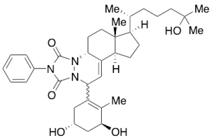
pre-Calcitriol PTAD Adduct (Mixture of Diastereomers)
$294.98 Add to cart View Product DetailsMolecular Formula : C35 H49 N3 O5
-
Procalcitonin (PCT) Antibody (PE9), mAb, Mouse
$127.65 Add to cart View Product DetailsPCT
is a 116 amino acid (aa) protein. It is comprised of three sections of a 57 aa
N-terminal PCT, a 32 aa calcitonin and a 21 aa katacalcin. Calcitonin is a
hormone, derived from PCT cleavage. PCT is a good diagnosis marker for
bacterial infection. Other diseases such as sepsis, inflammation, surgery, heat
shock, burn injuries and cardiogenic shock can also cause an increase of PCT
level in blood. -
Progesterone (5F2HC)
$230.29 Add to cart View Product DetailsThe
progesterone is a steroid hormone which is secreted by the female reproductive
systems. Its concentration is associated with the development and regression of
the corpus luteum. The progesterone is used in evaluation of ovulation status and
assessment of the luteal phase -
ProGRP Antibody (11B5), mAb, Mouse
$281.18 Add to cart View Product DetailsProgastrin-releasing peptide (ProGRP) is a stable
precursor of the gastrin releasing peptid. It is composed of GRP (residues
1–27), a cleavage site (residues 28–30), and a constant region (residues 31–98).
ProGRP is a tumor marker for the diagnosis and monitoring of small cell lung
cancer (SCLC). -
ProGRP Antibody (1C10), mAb, Mouse
$281.18 Add to cart View Product DetailsProgastrin-releasing peptide (ProGRP) is a stable
precursor of the gastrin releasing peptid. It is composed of GRP (residues
1–27), a cleavage site (residues 28–30), and a constant region (residues 31–98).
ProGRP is a tumor marker for the diagnosis and monitoring of small cell lung
cancer (SCLC). -
Prostaglandin E2
$332.94 Add to cart View Product DetailsProstaglandin E2
-
PSA (7H2), mAb, Mouse
$104.36 Add to cart View Product DetailsProstate specific antigen (PSA) is a serine protease produced by the prostate gland. In serum, it exists in different forms, cPSA, mainly including PSA-ACT (alpha-1-antichymotrypsin) and PSA-A2M (alpha-2-macroglobulin), fPSA (free PSA), etc. The blood level of PSA is increased in men with prostate cancer and it is considered as an indicator for prostate cancer, tumor recurrence and response to therapy.
-
PSA (8A12), mAb, Mouse
$104.36 Add to cart View Product DetailsProstate specific antigen (PSA) is a serine protease produced by the prostate gland. In serum, it exists in different forms, cPSA, mainly including PSA-ACT (alpha-1-antichymotrypsin) and PSA-A2M (alpha-2-macroglobulin), fPSA (free PSA), etc. The blood level of PSA is increased in men with prostate cancer and it is considered as an indicator for prostate cancer, tumor recurrence and response to therapy.
-

PSA Antibody (8A9B8), mAb, Mouse
$168.19 Add to cart View Product DetailsPSA monoclonal antibodies (5B10D4, 8A9B8, 1G9G8, 4A5D10 and 1A7D4) recognize total PSA, freePSA and ACT- PSA Complex (Prostate Specific Antigen / a-1 Antichymotrypsin Complex).
-
PSMA Fc Chimera, Human
$1,725.00 Add to cart View Product DetailsProstate-specific membrane antigen (PSMA) also known as Folate hydrolase 1 (FOLH1), Folylpoly-gamma-glutamate carboxypeptidase (FGCP), Glutamate carboxypeptidase 2 (GCP2), N-acetylated-alpha-linked acidic dipeptidase I (NAALAD1), is a type II membrane glycoprotein that is expressed in prostate tissue and to a lesser extent in the peripheral and central nervous system, small intestinal, and salivary gland tissues. PSMA has both folate hydrolase and N-acetylated-alpha-linked-acidic dipeptidase (NAALADase) activity and has a preference for tri-alpha-glutamate peptides. The catalytic activity of PSMA involves the release of unsubstituted C-terminal glutamyl residues, typically from Ac-Asp-Glu or folylpoly-gamma-glutamates. PSMA is used as a diagnostic and prognostic indicator of prostate cancer, and as a possible marker for various neurological disorders such as schizophrenia, Alzheimer’s disease, and Huntington’s disease.
-
PSMA, His, Human
$1,552.50 Add to cart View Product DetailsProstate-specific membrane antigen (PSMA) also known as Folate hydrolase 1 (FOLH1), Folylpoly-gamma-glutamate carboxypeptidase (FGCP), Glutamate carboxypeptidase 2 (GCP2), N-acetylated-alpha-linked acidic dipeptidase I (NAALAD1), is a type II membrane glycoprotein that is expressed in prostate tissue and to a lesser extent in the peripheral and central nervous system, small intestinal, and salivary gland tissues. PSMA has both folate hydrolase and N-acetylated-alpha-linked-acidic dipeptidase (NAALADase) activity and has a preference for tri-alpha-glutamate peptides. The catalytic activity of PSMA involves the release of unsubstituted C-terminal glutamyl residues, typically from Ac-Asp-Glu or folylpoly-gamma-glutamates. PSMA is used as a diagnostic and prognostic indicator of prostate cancer, and as a possible marker for various neurological disorders such as schizophrenia, Alzheimer’s disease, and Huntington’s disease.
-
PTH (1-84aa), Human
$819.38 Add to cart View Product DetailsParathyroid Hormone (PTH) is the most important endocrine regulator of calcium and phosphorus con- centration in extracellular fluid. Parathyroid Hormone (PTH) is secreted from cells of the parathyroid glands and finds Parathyroid Hormone’s major target cells in bone and kidney. Like most other protein hormones, Parathyroid Hormone (PTH) is synthesized as a preprohormone. After intracellular processing, the mature hormone is packaged within the Golgi into secretory vesicles, the secreted into blood by exocytosis. Parathyroid Hormone (PTH) is secreted as a linear protein of 84 amino acids. Recombinant Human Parathyroid Hormone (PTH) produced in E. coli is a single, non-glycosylated, polypeptide chain containing 84 amino acids.
-
PTH (7-34aa), Human
$1,138.50 Add to cart View Product DetailsPolypeptide hormones secreted by the parathyroid glands, which promote release of calcium from bone to extracellular fluid by activating osteoblasts and inhibiting osteoclasts, indirectly promote increased intestinal absorption of calcium, and promote renal tubular reabsorption of calcium and increased renal excretion of phosphates. It is a major regulator of bone metabolism. Secretion of parathyroid hormone increases when the level of calcium in the extracellular fluid is low. Its action is opposed by calcitonin.
-
PTH (8H4HC)
$319.13 Add to cart View Product Detailsbeta test product of PTH Antibody (8H4HC)
-
PTH (NT7HC)
$319.13 Add to cart View Product Detailsbeta test product of PTH Antibody (NT7HC)
-
PTH Antibody (M18-3), mAb, Mouse
$319.13 Add to cart View Product DetailsParathyroid
hormone (PTH) is synthesized by the parathyroid glands and secreted into the
blood stream. Intact PTH is a single polypeptide containing 84 amino acids.
PTH, which plays an important role in maintaining calcium homeostasis, is a
useful biomarker in the diagnosis of calcium related disorders. -
PTH Antibody (NT7M), mAb, Mouse
$319.13 Add to cart View Product DetailsParathyroid
hormone (PTH) is synthesized by the parathyroid glands and secreted into the
blood stream. Intact PTH is a single polypeptide containing 84 amino acids.
PTH, which plays an important role in maintaining calcium homeostasis, is a
useful biomarker in the diagnosis of calcium related disorders.