1mg
Showing 3801–3850 of 4152 results
-
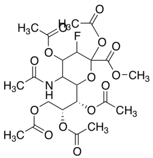
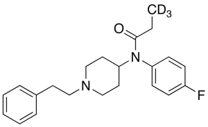
O-Desmethyl Indomethacin-d4
$264.79 Add to cart View Product DetailsMolecular Formula : C18H10D4ClNO4
-
O-Desmethyl Mebeverine Acid
$114.71 Add to cart View Product DetailsMolecular Formula : C15 H23 N O3
-

O-Desmethyl Mebeverine Acid O-Beta-D-Glucuronide Ammonium Salt
$166.46 Add to cart View Product DetailsMolecular Formula : C21H31NO9 xNH3
-
O-Desmethyl Metoprolol-d5
$185.44 Add to cart View Product DetailsMolecular Formula : C14H18D5NO3
-

O-Desmethyl Midostaurin
$251.85 Add to cart View Product DetailsMolecular Formula : C34 H28 N4 O4
-
O-Desmethyl Midostaurin-13C6
$1,144.54 Add to cart View Product DetailsMolecular Formula : C2813C6H28N4O4
-
O-Desmethyl Mycophenolic Acid
$206.14 Add to cart View Product DetailsMolecular Formula : C16 H18 O6
-

O-Desmethyl Quinine
$67.28 Add to cart View Product DetailsMolecular Formula : C19 H22 N2 O2
-
O-Desmethyl Tramadol Beta-D-Glucuronide (Mixture of Diastereomers)
$263.93 Add to cart View Product DetailsMolecular Formula : C21 H31 N O8
-

O-Desmethyl Tramadol-d6
$190.61 Add to cart View Product DetailsMolecular Formula : C15 2H6 H17 N O2
-
O-Desmethyl Venlafaxine Cyclic Impurity
$280.31 Add to cart View Product DetailsMolecular Formula : C16 H23 N O2
-
O-tert-Butyl-2-hydroxy Efavirenz-d5
$192.34 Add to cart View Product DetailsMolecular Formula : C18 D5 H14 Cl F3 N O3
-

O,O-Diacetyl-O6-demethylsalutaridine
$166.46 Add to cart View Product DetailsMolecular Formula : C22H23NO6
-
O,O-Diethyl Dithiophosphate-13C4 Ammonium Salt
$213.04 Add to cart View Product DetailsMolecular Formula : 13C4H14NO2PS2
-

o,p’-Dicofol
$95.74 Add to cart View Product DetailsMolecular Formula : C14 H9 Cl5 O
-

O4-Desacetyl Vinblastinic Acid
$171.64 Add to cart View Product DetailsMolecular Formula : C43H54N4O8
-
O4-Ethylthymine-d5
$64.69 Add to cart View Product DetailsMolecular Formula : C7 D5 H7 N2 O2
-

Omega-Hydroxy Fentanyl-d5
$214.76 Add to cart View Product DetailsMolecular Formula : C22H23D5N2O2
-
OSM (209aa), Human
$2,018.25 Add to cart View Product DetailsOncostatin M (OSM) is a multifunctional cytokine, and belongs to Interleukin-6 (IL-6) subfamily, which also includes IL-11, leukemia inhibitory factor (LIF), ciliary neurotropic factor, cardiotrophin-1, and novel neurotropin-1. In vivo, OSM is secreted from activated T cells, monocytes, neutrophils, and endothelial cells. OSM is related to LIF, and shares a receptor with LIF in human. Human OSM can bind to gp130 and recruit OSM Receptor β or LIF Receptor β to form a ternary complex. OSM stimulates the growth of different types of cells, including megakaryocytes, fibroblasts, vascular endothelial cells, and T cells. OSM inhibits the proliferation of several cancer cell lines, such as solid tissue tumor cells, lung cancer cells, melanoma cells, and breast cancer cells.
-
OSM (227aa), Human
$2,018.25 Add to cart View Product DetailsOncostatin M (OSM) is a multifunctional cytokine, and belongs to Interleukin-6 (IL-6) subfamily, including IL-11, leukemia inhibitory factor (LIF), ciliary neurotropic factor, cardiotrophin-1, and novel neurotropin-1. In vivo, OSM is secreted from activated T cells, monocytes, neutrophils, and endothelial cells. OSM is related to LIF, and share a receptor with LIF in human. Human OSM can bind to gp130 and recruit OSM Receptor β or LIF Receptor β to form a ternary complex. OSM stimulates the growth of different types of cells, including megakaryocytes, fibroblasts, vascular endothelial cells, and T cells. On the other hand, OSM inhibits the proliferation of several cancer cell lines, such as solid tissue tumor cells, lung cancer cells, melanoma cells, and breast cancer cells.
-
OSM, Mouse
$2,018.25 Add to cart View Product DetailsOncostatin M (OSM) is a multifunctional cytokine, and belongs to Interleukin-6 (IL-6) subfamily, which also includes IL-11, leukemia inhibitory factor (LIF), ciliary neurotropic factor, cardiotrophin-1, and novel neurotropin-1. In vivo, OSM is secreted from activated T cells, monocytes, neutrophils, and endothelial cells. OSM is related to LIF, and shares a receptor with LIF in human. Human OSM can bind to gp130 and recruit OSM Receptor β or LIF Receptor β to form a ternary complex. OSM stimulates the growth of different types of cells, including megakaryocytes, fibroblasts, vascular endothelial cells, and T cells. OSM inhibits the proliferation of several cancer cell lines, such as solid tissue tumor cells, lung cancer cells, melanoma cells, and breast cancer cells.
-
OSM, Mouse(HEK 293-expressed)
$2,018.25 Add to cart View Product DetailsOncostatin M (OSM) is a multifunctional cytokine, and belongs to Interleukin-6 (IL-6) subfamily, which also includes IL-11, leukemia inhibitory factor (LIF), ciliary neurotropic factor, cardiotrophin-1, and novel neurotropin-1. In vivo, OSM is secreted from activated T cells, monocytes, neutrophils, and endothelial cells. OSM is related to LIF, and shares a receptor with LIF in human. Human OSM can bind to gp130 and recruit OSM Receptor β or LIF Receptor β to form a ternary complex. OSM stimulates the growth of different types of cells, including megakaryocytes, fibroblasts, vascular endothelial cells, and T cells. OSM inhibits the proliferation of several cancer cell lines, such as solid tissue tumor cells, lung cancer cells, melanoma cells, and breast cancer cells.
-

OX40 Fc Chimera, Mouse
$1,035.00 Add to cart View Product DetailsOX40 (TNFRSF4, CD134) is a member of the tumor necrosis factor (TNF) receptor superfamily that regulates T cell activity and immune responses. The OX40 protein contains four cysteine rich domains, a transmembrane domain, and a cytoplasmic tail containing a QEE motif. OX40 is primarily expressed on activated CD4+ and CD8+ T-cells, while the OX40 ligand (OX40L, TNFSF4, CD252) is predominantly expressed on activated antigen presenting cells. The engagement of OX40 with OX40L leads to the recruitment of TNF receptor-associated factors (TRAFs) and results in the formation of a TCR-independent signaling complex. One component of this complex, PKCθ, activates the NF-κB pathway. OX40 signaling through Akt can also enhance TCR signaling directly. Research studies indicate that the OX40L-OX40 pathway is associated with inflammation and autoimmune diseases. Additional research studies show that OX40 agonists augment anti-tumor immunity in several cancer types.
-

OX40/TNFRSF4, His, Human
$1,035.00 Add to cart View Product DetailsOX40 is a T cell co-stimulatory molecule of the TNF receptor superfamily that coordinates with other co-stimulators (CD28, CD40, CD30, CD27 and 4-1BB) to manage the activation of the immune response. Tumor necrosis factor ligand superfamily member 4 (TNFSF4) is also known as glycoprotein Gp34, OX40 ligand (OX40L), TAX transcriptionally-activated glycoprotein 1 and CD252, which belongs to the tumor necrosis factor family. TNFSF4 is the ligand for CD134 and is expressed on cells such as DC2s (a subtype of dendritic cells) enabling amplification of Th2 cell differentiation. OX40 is constitutively expressed on Tregs and enhances the sensitivity of Tregs to IL-2, thus promoting Treg proliferation. OX40 has also shown to decrease the cells’ immunosuppressive activity on effector T cells. OX40-OX40 Ligand signaling is involved in allergic airway inflammation, graft-versus-host disease and autoimmune disease.
-
![P-[(3-Bromo-7-cyano-2-naphthalenyl)difluoromethyl]phosphonic Acid Diammonium Salt](https://advatechgroup.com/wp-content/uploads/sys-masterimagesh3ch6b10621080371230B682735-300x90.png)
P-[(3-Bromo-7-cyano-2-naphthalenyl)difluoromethyl]phosphonic Acid Diammonium Salt
$283.76 Add to cart View Product DetailsMolecular Formula : C12H13BrF2N3O3P
-
P-3FAX-Neu5Ac
$91.43 Add to cart View Product DetailsMolecular Formula : C22H30FNO14
-

p-Chlorobenzyl Bromide-d6
$166.46 Add to cart View Product DetailsMolecular Formula : C7 D6 Br Cl
-
p-Cresol Rutinoside
$224.25 Add to cart View Product DetailsMolecular Formula : C19 H28 O10
-
p-Cresol Rutinoside-d7 (major)
$179.40 Add to cart View Product DetailsMolecular Formula : C19 D7 H21 O10
-
p-Cresol-d4
$194.93 Add to cart View Product DetailsMolecular Formula : C7 D4 H4 O
-
p-Fluoro Fentanyl-d3
$237.19 Add to cart View Product DetailsMolecular Formula : C22H24D3FN2O
-
p-Hydroxybenzoylecgonine
$165.60 Add to cart View Product DetailsMolecular Formula : C16 H19 N O5
-
p-Hydroxymephentermine Hydrochloride
$176.81 Add to cart View Product DetailsMolecular Formula : C11 H17 N O . Cl H
-
p-O-Desmethyl Verapamil
$132.83 Add to cart View Product DetailsMolecular Formula : C26 H36 N2 O4
-
p-Tau181 Antibody(A2F), mAb, Mouse
$380.36 Add to cart View Product DetailsMicrotubule associated protein tau is a neuronal microtubule-associated protein found predominantly in brain. Abnormal phosphorylation of tau is involved in the pathogenesis of Alzheimer’s disease (AD). The tau protein phosphorylated at threonine 181 (p-tau181) is a promising diagnostic biomarker for AD.
-
PAPP-A (19D1), mAb, Mouse
$116.44 Add to cart View Product DetailsPregnancy-associated plasma protein A (PAPP-A) is a high molecular weight protein. htPAPP-A is a protein complex which comprises of two PAPP-A subunits and two proform of eosinophil major basic protein. It exists in the blood of pregnant woman. dPAPP-A is a htPAPP-A dimmer which is composed of two PAPP-A subunits. htPAPP-A serves as a useful marker for the diagnosis of Down syndrome (DS).
-
PAPP-A (1AC2), mAb, Mouse
$116.44 Add to cart View Product DetailsPregnancy-associated plasma protein A (PAPP-A) is a high molecular weight protein. htPAPP-A is a protein complex which comprises of two PAPP-A subunits and two proform of eosinophil major basic protein. It exists in the blood of pregnant woman. dPAPP-A is a htPAPP-A dimmer which is composed of two PAPP-A subunits. htPAPP-A serves as a useful marker for the diagnosis of Down syndrome (DS).
-
PCT (16A1), mAb, Mouse
$131.10 Add to cart View Product DetailsPCT, a 116 amino acid (aa) protein, is comprised of three sections including a 57 aa N-terminal PCT, a 32 aa calcitonin and a 21 aa katacalcin. Calcitonin is a hormone, derived from PCT cleavage. PCT is a good diagnosis marker for bacterial infection. Other diseases such as sepsis, inflammation, surgery, heat shock, burn injuries and cardiogenic shock can also cause an increase of PCT level in blood.
-
PCT (18D2), mAb, Mouse
$131.10 Add to cart View Product DetailsPCT, a 116 amino acid (aa) protein, is comprised of three sections including a 57 aa N-terminal PCT, a 32 aa calcitonin and a 21 aa katacalcin. Calcitonin is a hormone, derived from PCT cleavage. PCT is a good diagnosis marker for bacterial infection. Other diseases such as sepsis, inflammation, surgery, heat shock, burn injuries and cardiogenic shock can also cause an increase of PCT level in blood.
-
PCT (18M3), mAb, Mouse
$131.10 Add to cart View Product DetailsPCT, a 116 amino acid (aa) protein, is comprised of three sections including a 57 aa N-terminal PCT, a 32 aa calcitonin and a 21 aa katacalcin. Calcitonin is a hormone, derived from PCT cleavage. PCT is a good diagnosis marker for bacterial infection. Other diseases such as sepsis, inflammation, surgery, heat shock, burn injuries and cardiogenic shock can also cause an increase of PCT level in blood.
-
PCT (PE21), mAb, Mouse
$131.10 Add to cart View Product DetailsPCT, a 116 amino acid (aa) protein, is comprised of three sections including a 57 aa N-terminal PCT, a 32 aa calcitonin and a 21 aa katacalcin. Calcitonin is a hormone, derived from PCT cleavage. PCT is a good diagnosis marker for bacterial infection. Other diseases such as sepsis, inflammation, surgery, heat shock, burn injuries and cardiogenic shock can also cause an increase of PCT level in blood.
-
PD-1 (2E5), mAb, Mouse
$1,306.69 Add to cart View Product DetailsProgrammed Cell Death Protein 1 (PD-1), is cell surface receptor expressing on T cells and pro-B cells. The binding of PD-1 to its two ligands, PD-L1 and PD-L2, could result in down-regulation of the immune system by inhibiting the T-cell activation process. Thus, PD-1 is an important immune checkpoint and a popular target for therapeutic antibodies against many cancers.
-

PD-1 Fc Chimera, Human
$1,035.00 Add to cart View Product DetailsProgrammed cell death protein 1, also known as PD-1 and CD279 (cluster of differentiation 279) or PDCD1, is a protein that in humans is encoded by the PDCD1 gene. PD-1 is a cell surface receptor that belongs to the immunoglobulin superfamily and is expressed on T cells and pro-B cells.PD-1 binds two ligands, PD-L1 and PD-L2. PD-1 and its ligands play an important role in down regulating the immune system by preventing the activation of T-cells, which in turn reduces autoimmunity and promotes self-tolerance. The inhibitory effect of PD-1 is accomplished through a dual mechanism of promoting apoptosis (programmed cell death) in antigen specific T-cells in lymph nodes while simultaneously reducing apoptosis in regulatory T cells (suppressor T cells)
-

PD-1 Fc Chimera, Mouse
$1,035.00 Add to cart View Product DetailsProgrammed cell death protein 1, also known as PD-1 and CD279 (cluster of differentiation 279) or PDCD1, is a protein that in humans is encoded by the PDCD1 gene. PD-1 is a cell surface receptor that belongs to the immunoglobulin superfamily and is expressed on T cells and pro-B cells.PD-1 binds two ligands, PD-L1 and PD-L2. PD-1 and its ligands play an important role in down regulating the immune system by preventing the activation of T-cells, which in turn reduces autoimmunity and promotes self-tolerance. The inhibitory effect of PD-1 is accomplished through a dual mechanism of promoting apoptosis (programmed cell death) in antigen specific T-cells in lymph nodes while simultaneously reducing apoptosis in regulatory T cells (suppressor T cells).
-
PD-1, His, Human
$1,293.75 Add to cart View Product DetailsProgrammed death (PD-1) is an immunoinhibitory receptor that belongs to the CD28 family and is expressed on T cells, B cells, monocytes, natural killer cells, and many tumor-infiltrating lymphocytes (TILs); PD-1 is a type I membrane protein of 268 amino acids and which structure includes an extracellular IgV domain followed by a transmembrane region and an intracellular tail. The intracellular tail contains two phosphorylation sites located in an immunoreceptor tyrosine-based inhibitory motif and an immunoreceptor tyrosine-based switch motif, which suggests that PD-1 negatively regulates TCR signals. This is consistent with binding of SHP-1 and SHP-2 phosphatases to the cytoplasmic tail of PD-1 upon ligand binding. It has 2 ligands that have been described PD-L1(B7H1) and PD-L2(B7-DC); PD-1 induction on activated T cells occurs in response to PD-L1 or L2 engagement and limits effector T-cell activity in peripheral organs and tissues during inflammation, thus preventing autoimmunity.Recombinant Human PD-1 produced in HEK293 cells is a polypeptide chain containing 149 amino acids with C-terminal 6×His. A fully biologically active molecule, rhPD-1 has a molecular mass of 30-40 kDa analyzed by reducing SDS-PAGE and is obtained by chromatographic techniques at GenScript.
-
PD-L1 Fc Chimera, Human
$1,035.00 Add to cart View Product DetailsProgrammed death-ligand 1 (PD-L1) also known as cluster of differentiation 274 (CD274) or B7 homolog 1 (B7-H1), is a protein that in humans is encoded by the CD274 gene. PD-L1 is a 40 kDa type 1 transmembrane protein that has been speculated to play a major role in suppressing the immune system during particular events such as pregnancy, tissue allografts, autoimmune disease and other disease states such as hepatitis. Normally the immune system reacts to foreign antigens where there is some accumulation in the lymph nodes or spleen which triggers a proliferation of antigen-specific CD8+ T cell. The formation of PD-1 receptor / PD-L1 or B7.1 receptor /PD-L1 ligand complex transmits an inhibitory signal which reduces the proliferation of these CD8+ T cells at the lymph nodes and supplementary to that PD-1 is also able to control the accumulation of foreign antigen specific T cells in the lymph nodes through apoptosis which is further mediated by a lower regulation of the gene Bcl-2. PD-L1 binds to its receptor, PD-1, found on activated T cells, B cells, and myeloid cells, to modulate activation or inhibition. Recombinant Human PD-L1(B7-H1) Fc Chimera produced in CHO cells is a polypeptide chain containing 457 amino acids. A fully biologically active molecule, rh PD‑L1(B7-H1) has a molecular mass of 70-72 kDa analyzed by reducing SDS-PAGE and is obtained by chromatographic techniques at GenScript.
-
PD-L1 Fc Chimera, Mouse
$1,035.00 Add to cart View Product DetailsProgrammed death-ligand 1 (PD-L1) also known as cluster of differentiation 274 (CD274) or B7 homolog 1 (B7-H1), is a protein that in humans is encoded by the CD274 gene. PD-L1 is a 40 kDa type 1 transmembrane protein that has been speculated to play a major role in suppressing the immune system during particular events such as pregnancy, tissue allografts, autoimmune disease and other disease states such as hepatitis. Normally the immune system reacts to foreign antigens where there is some accumulation in the lymph nodes or spleen which triggers a proliferation of antigen-specific CD8+ T cell. The formation of PD-1 receptor / PD-L1 or B7.1 receptor /PD-L1 ligand complex transmits an inhibitory signal which reduces the proliferation of these CD8+ T cells at the lymph nodes and supplementary to that PD-1 is also able to control the accumulation of foreign antigen specific T cells in the lymph nodes through apoptosis which is further mediated by a lower regulation of the gene Bcl-2. PD-L1 binds to its receptor, PD-1, found on activated T cells, B cells, and myeloid cells, to modulate activation or inhibition.
-

PD-L1, His, Human
$1,035.00 Add to cart View Product DetailsProgrammed death-ligand 1 (PD-L1) also known as cluster of differentiation 274 (CD274) or B7 homolog 1 (B7-H1), is a protein that in humans is encoded by the CD274 gene. PD-L1 is a 40 kDa type 1 transmembrane protein that has been speculated to play a major role in suppressing the immune system during particular events such as pregnancy, tissue allografts, autoimmune disease and other disease states such as hepatitis. Normally the immune system reacts to foreign antigens where there is some accumulation in the lymph nodes or spleen which triggers a proliferation of antigen-specific CD8+ T cell. The formation of PD-1 receptor / PD-L1 or B7.1 receptor /PD-L1 ligand complex transmits an inhibitory signal which reduces the proliferation of these CD8+ T cells at the lymph nodes and supplementary to that PD-1 is also able to control the accumulation of foreign antigen specific T cells in the lymph nodes through apoptosis which is further mediated by a lower regulation of the gene Bcl-2. PD-L1 binds to its receptor, PD-1, found on activated T cells, B cells, and myeloid cells, to modulate activation or inhibition. Recombinant Human PD-L1(B7-H1) Fc Chimera produced in CHO cells is a polypeptide chain containing 457 amino acids. A fully biologically active molecule, rh PD‑L1(B7-H1) has a molecular mass of 70-72 kDa analyzed by reducing SDS-PAGE and is obtained by chromatographic techniques at GenScript.
-

PD-L2 Fc Chimera, Human
$1,035.00 Add to cart View Product DetailsPD-L1 and PD-L2 are ligands for PD-1, a costimulatory molecule that plays an inhibitory role in regulating T cell activation in the periphery. PD-L2 also known as PD-L2, B7-DC serves as a negative and a positive regulator of T cell function. The expression and function of PD-L2 are similar to PD-L1. Both PD-L2−PD-1 and PD-L1−PD-1 signals inhibit T cell proliferation by blocking cell cycle progression but not by increasing cell death. PD-L2−PD-1 interactions are able to inhibit TCR-mediated proliferation and cytokine production in the absence of CD28 costimulation. Threshold for T cell activation may be a balance between activating signals, such as those delivered by the engagement of CD28 by B7-1 and B7-2, and inhibitory signals, mediated by engagement of PD-1 by PD-L1 and PD-L2. The structural conservation of B7-like and CD28-like receptors may reflect the distance between T cells and APCs in the immunological synapse. The PD-L−PD-1 pathway may play a key role in the induction and/or maintenance of peripheral tolerance and autoimmune disease. Because PD-L1 and PD-L2 can inhibit effector T cell proliferation and cytokine production, the PD-L−PD-1 pathway may be an attractive therapeutic target. Blocking the PD-1 pathway may enhance anti-tumor immunity, whereas stimulating this pathway may be useful for down-regulating ongoing immune responses in transplant rejection and autoimmune and allergic diseases.
-

PDGF-AA, Mouse
$1,712.06 Add to cart View Product DetailsPlatelet-Derived Growth Factor-AA (PDGF-AA) is one of five dimers (PDGF-AA, AB, BB, CC, and DD) formed by 4 different PDGF subunits. In chemical terms, platelet-derived growth factor is a dimeric glycoprotein composed of two A (-AA) or two B (-BB) chains or a combination of the two (-AB). The dimeric isoforms PDGFAA, AB and BB are differentially expressed in various cell types, and their effects are mediated through two distinct receptors termed α and β. Differences exist in isoform binding to each receptor. Ingeneral, PDGF isoforms are potent mitogens for connective tissue cells including dermal fibroblasts, glial cells, arterial smooth muscle cells and some epithelial andendothelial cells. In addition to its activity as a mitogen, PDGF is chemotactic for fibroblasts, smooth muscle cells, neutrophils and mononuclear cells. PDGF-AA plays a significant role in blood vessel formation (angiogenesis).