50g
Showing 1601–1650 of 1859 results
-
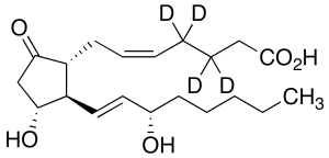
Prostaglandin E2-d4
$90.56 Add to cart View Product DetailsMolecular Formula : C20H28D4O5
-

PSMA Fc Chimera, Human
$185.44 Add to cart View Product DetailsProstate-specific membrane antigen (PSMA) also known as Folate hydrolase 1 (FOLH1), Folylpoly-gamma-glutamate carboxypeptidase (FGCP), Glutamate carboxypeptidase 2 (GCP2), N-acetylated-alpha-linked acidic dipeptidase I (NAALAD1), is a type II membrane glycoprotein that is expressed in prostate tissue and to a lesser extent in the peripheral and central nervous system, small intestinal, and salivary gland tissues. PSMA has both folate hydrolase and N-acetylated-alpha-linked-acidic dipeptidase (NAALADase) activity and has a preference for tri-alpha-glutamate peptides. The catalytic activity of PSMA involves the release of unsubstituted C-terminal glutamyl residues, typically from Ac-Asp-Glu or folylpoly-gamma-glutamates. PSMA is used as a diagnostic and prognostic indicator of prostate cancer, and as a possible marker for various neurological disorders such as schizophrenia, Alzheimer’s disease, and Huntington’s disease.
-
PSMA, His, Human
$168.19 Add to cart View Product DetailsProstate-specific membrane antigen (PSMA) also known as Folate hydrolase 1 (FOLH1), Folylpoly-gamma-glutamate carboxypeptidase (FGCP), Glutamate carboxypeptidase 2 (GCP2), N-acetylated-alpha-linked acidic dipeptidase I (NAALAD1), is a type II membrane glycoprotein that is expressed in prostate tissue and to a lesser extent in the peripheral and central nervous system, small intestinal, and salivary gland tissues. PSMA has both folate hydrolase and N-acetylated-alpha-linked-acidic dipeptidase (NAALADase) activity and has a preference for tri-alpha-glutamate peptides. The catalytic activity of PSMA involves the release of unsubstituted C-terminal glutamyl residues, typically from Ac-Asp-Glu or folylpoly-gamma-glutamates. PSMA is used as a diagnostic and prognostic indicator of prostate cancer, and as a possible marker for various neurological disorders such as schizophrenia, Alzheimer’s disease, and Huntington’s disease.
-
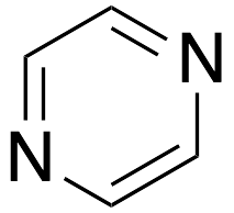
Pyrazine
$276.86 Add to cart View Product DetailsMolecular Formula : C4 H4 N2
-

Pyridine-Sulfur Trioxide Complex
$62.10 Add to cart View Product DetailsMolecular Formula : C5H5N.SO3
-

Pyridinium Dichromate
$87.11 Add to cart View Product DetailsMolecular Formula : C10H12Cr2N2O7
-
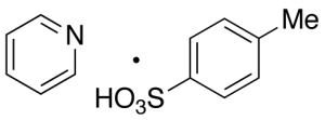
Pyridinium p-Toluenesulfonate
$130.24 Add to cart View Product DetailsMolecular Formula : C12H13NO3S
-
Pyrrole-1-carboxylic Acid tert-Butyl Ester
$187.16 Add to cart View Product DetailsMolecular Formula : C9H13NO2
-

Pyrrole-2-carboxylic Acid
$633.08 Add to cart View Product DetailsMolecular Formula : C5H5NO2
-

Pyruvic Acid
$81.94 Add to cart View Product DetailsMolecular Formula : C3 H4 O3
-

Quinidine Hydrochloride Dihydrate
$223.39 Add to cart View Product DetailsMolecular Formula : C20 H24 N2 O2 . Cl H . 2 H2 O
-

Quinidine Sulfate (>85%)
$357.94 Add to cart View Product DetailsMolecular Formula : C20 H24 N2 O2 . H2 O4 S
-
Quinidine Sulfate, Dihydrate, USP
$316.37 Add to cart View Product DetailsQuinidine Sulfate, Dihydrate, USP
-
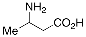
rac 3-Aminobutyric Acid
$791.78 Add to cart View Product DetailsMolecular Formula : C4 H9 N O2
-
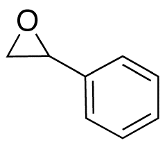
rac Styrene Oxide
$66.41 Add to cart View Product DetailsMolecular Formula : C8H8O
-

rac-Citronellal (>90%)
$177.68 Add to cart View Product DetailsMolecular Formula : C10 H18 O
-

rac-Gamma-Nonanolactone
$155.25 Add to cart View Product DetailsMolecular Formula : C9 H16 O2
-

Rapidosept
$137.14 Add to cart View Product DetailsMolecular Formula : C7 H6 Cl2 O
-
RBP4, His, Human
$155.25 Add to cart View Product DetailsThe properties of retinol binding protein is the transport carrier of vitamin A in the plasma. Human-retinol binding protein is a single-chain polypeptide with a molecular weight of approximately 21000 and one binding site for retinol and other forms of vitamin A. In addition, compounds related to retinol, such as retinal, retinoic acid, retinyl esters and geometric isomers of retinol and of retinal were evaluated for their ability to bind to this protein. In plasma, RBP4-retinol forms a complex with transthyretin (TTR), also known as thyroxine-binding protein and prealbumin. Defects in RBP4 cause retinol-binding protein deficiency, which affects night vision.
-
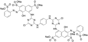
Reactive Red 120 Sodium Salt (Technical Grade)
$130.24 Add to cart View Product DetailsMolecular Formula : C44H24Cl2N14Na6O20S6
-

Rochelle’s Salt
$227.70 Add to cart View Product DetailsMolecular Formula : C4 H4 O6 . K . Na . 4 H2 O
-
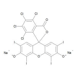
Rose Bengal Sodium Salt, Technical Grade
$244.95 Add to cart View Product DetailsMolecular Formula : C20 H2 Cl4 I4 O5 . 2 Na
-

RSPO1, His, Human
$452.81 Add to cart View Product DetailsRSPO1 is a secreted protein, containing 2 FU(furin-like) repeats and 1 TSP type-1 domain and belonging to the R-spondin family. RSPO1 is required for the early development of gonads, regardless of sex. It has been found in mice only eleven days after fertilization. To induce cell proliferation, it acts synergistically with WNT4. They help stabilize β catenin, which activates downstream targets. RSPO1 is necessary in female sex development. It augments the WNT/β catenin pathway to oppose male sex development. In critical gonadal stages, between six and nine weeks after fertilization, the ovaries upregulate it while the testes downregulate it. RSPO1 can potentially aid in the treatment of mucositis, which is characterized by inflammation of the oral cavity. This unfortunate condition often accompanies chemotherapy and radiation in cancer patients with head and neck tumors.
-

S100A1, His, Human
$392.44 Add to cart View Product DetailsS100 calcium-binding protein A1 (S100A1) is a small calcium binding protein with EF-hand structures and belongs to the S100 family. S100 proteins include at least 25 members which are located as a cluster on human chromosome 1q21. S100A1 is found in the heart, skeletal muscle, brain, and kidney.S100A1 mainly resides on the sarcoplasmic reticulum, mitochondria and myofilaments. S100A1 may function in stimulation of Ca2+ induced Ca2+ release, inhibition of microtubule assembly, and inhibition of protein kinase C-mediated phosphorylation. Reduced expression of this protein has been implicated in cardiomyopathies.
-

Saccharin
$120.75 Add to cart View Product DetailsMolecular Formula : C7 H5 N O3 S
-
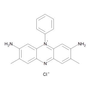
Safranin T (>75% Purity)
$319.99 Add to cart View Product DetailsMolecular Formula : C20 H19 N4 . Cl
-
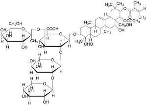
Saponin (Technical Grade)
$77.63 Add to cart View Product DetailsMolecular Formula : C62H96O27
-
sCD40L, Human
$155.25 Add to cart View Product DetailsCD40 ligand, CD40L (also known as CD154, TRAP or gp39), is a 261 amino acid type II transmembrane glycoprotein belonging to the TNF family, CD40L is expressed predominantly on activated CD4+ T lymphocytes, and also found in other types of cells, like NK cells, mast cells, basophils and eosinophils. Human CD40L shares 78% amino acid identity with its mouse counterpart. The receptor of CD40L is CD40, a type I transmembrane glycoprotein belonging to the TNF receptor family. CD40 is expressed on B lymphocytes, monocytes, dendritic cells and thymic epithelium. Although all monomeric, dimeric and trimeric forms of soluble CD40L can bind to CD40, the trimeric form of soluble CD40L has the most potent biological activity through oligomerization of cell surface CD40, a common feature of TNF receptor family members. CD40L mediates a range of activities on B cells including induction of activation-associated surface antigen, entry into cell cycle, isotype switching and Ig secretion and memory generation. CD40-CD40L interaction also plays important roles in monocyte activation and dendritic cell maturation.
-
SCF, Human (P. pastoris-expressed)
$155.25 Add to cart View Product DetailsStem cell factor (also known as SCF, KIT-ligand, KL, or steel factor) is a cytokine that binds to the c-KIT receptor (CD117). SCF can exist both as a transmembrane protein and a soluble protein. It stimulates the proliferation of myeloid, erythroid, and lymphoid progenitors in bone marrow cultures and has been shown to act synergistically with colony stimulating factors. SCF plays an important role in the hematopoiesis during embryonic development. SCF can regulates HSCs in the stem cell niche in the bone marrow. SCF has been shown to increase the survival of HSCs in vitro and contributes to the self-renewal and maintenance of HSCs in-vivo.
-
SCF, Mouse
$155.25 Add to cart View Product DetailsStem cell factor (also known as SCF, KIT-ligand, KL, or steel factor) is a cytokine that binds to the c-KIT receptor (CD117). SCF can exist both as a transmembrane protein and a soluble protein. It stimulates the proliferation of myeloid, erythroid, and lymphoid progenitors in bone marrow cultures and has been shown to act synergistically with colony stimulating factors. SCF plays an important role in the hematopoiesis during embryonic development. SCF can regulates HSCs in the stem cell niche in the bone marrow. SCF has been shown to increase the survival of HSCs in vitro and contributes to the self-renewal and maintenance of HSCs in-vivo.
-
SCF, Rat (HEK 293-expressed)
$155.25 Add to cart View Product DetailsStem cell factor (also known as SCF, KIT-ligand, KL, or steel factor) is a cytokine that binds to the c-KIT receptor (CD117). SCF can exist both as a transmembrane protein and a soluble protein. It stimulates the proliferation of myeloid, erythroid, and lymphoid progenitors in bone marrow cultures and has been shown to act synergistically with colony stimulating factors. SCF plays an important role in the hematopoiesis during embryonic development. SCF can regulates HSCs in the stem cell niche in the bone marrow. SCF has been shown to increase the survival of HSCs in vitro and contributes to the self-renewal and maintenance of HSCs in-vivo.
-
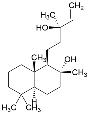
Sclareol (~90%)
$324.30 Add to cart View Product DetailsMolecular Formula : C20 H36 O2
-
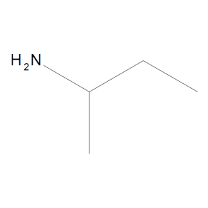
sec-Butylamine
$65.55 Add to cart View Product DetailsMolecular Formula : C4 H11 N
-

sec-Butylbenzene
$225.98 Add to cart View Product DetailsMolecular Formula : C10 H14
-
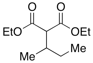
sec-Butylmalonic Acid Diethyl Ester
$3,132.60 Add to cart View Product DetailsMolecular Formula : C11H20O4
-
Selenious Acid, Reagent
$344.23 Add to cart View Product DetailsSelenious Acid, Reagent
-

Selenium(IV) Oxide
$154.39 Add to cart View Product DetailsMolecular Formula : No Data Available
-

Sephadex LH-20
$656.36 Add to cart View Product DetailsMolecular Formula : No Data Available
-
Shh (C24II), Human
$155.25 Add to cart View Product DetailsSonic Hedgehog (Shh) is a member of the Hedgehog (Hh) family of highly conserved proteins which are widely represented throughout the animal kingdom. In mammal, there are three related Hh proteins, Sonic (Shh), Desert (Dhh) and Indian (Ihh). They share a high degree of amino-acid sequence identity (e.g., Shh and Ihh are 93% identical). Sonic Hedgehog plays a role in cell growth, cell specialization, and the normal shaping (patterning) of the body. Shh is also important for development of the brain and spinal cord (central nervous system), eyes, limbs, and many other parts of the body.
-
Shh (C25II), Mouse
$155.25 Add to cart View Product DetailsSonic Hedgehog (Shh) is a member of the Hedgehog (Hh) family of highly conserved proteins which are widely represented throughout the animal kingdom. In mammal, there are three related Hh proteins, Sonic (Shh), Desert (Dhh) and Indian (Ihh). They share a high degree of amino-acid sequence identity (e.g., Shh and Ihh are 93% identical). Sonic Hedgehog plays a role in cell growth, cell specialization, and the normal shaping (patterning) of the body. Shh is also important for development of the brain and spinal cord (central nervous system), eyes, limbs, and many other parts of the body.
-

Shh, Mouse
$155.25 Add to cart View Product DetailsMembers of the Hedgehog (Hh) family are highly conserved proteins which are widely represented throughout the animal kingdom. The three known mammalian Hh proteins, Sonic (Shh), Desert (Dhh) and Indian (Ihh) are structurally related and share a high degree of amino-acid sequence identity (e.g., Shh and Ihh are 93% identical). The biologically active form of Hh molecules is obtained by autocatalytic cleavage of their precursor proteins and corresponds to approximately the N-terminal one half of the precursor molecule. Although Hh proteins have unique expression patterns and distinct biological roles within their respective regions of secretion, they use the same signaling pathway and can substitute for each other in experimental systems.
-
Siglec-10 Fc Chimera, Human
$137.14 Add to cart View Product DetailsSiglec-10 is immune system-restricted and highly expressed in peripheral blood leukocytes. Siglec-10 preferably binds to α-2,3- or α-2,6-linked sialic acid (similarity). Siglec10 is involved in the negative regulation of B cell antigen receptor signal transduction. The inhibition of B cell activation depends on PTPN6/SHP-1 (by similarity). The binding of Siglec10 to CD24 may be involved in the selective suppression of the immune response (by similarity) to risk-related molecular patterns (DAMPs) (such as HMGB1, HSP70 and HSP90). The combination of Siglec10 and CD24 may regulate the immune response of natural killer (NK) cells. Play a role in controlling autoimmunity (by similarity). In the process of initiating an adaptive immune response by CD8-α+ dendritic cells, cross-presentation is inhibited by weakening the formation of MHC class I peptide complexes. The function seems to imply the recruitment of PTPN6/SHP-1, which dephosphorylates NCF1 of the NADPH oxidase complex, thereby promoting phagosome acidification (by similarity).
-
Siglec-15 Fc Chimera, Human
$137.14 Add to cart View Product DetailsSiglec-15 preferentially recognizes the Neu5Acalpha2-6GalNAcalpha- structure. Siglec-15 associates with the activating adaptor proteins DNAX activation protein (DAP)12 and DAP10 via its lysine residue in the transmembrane domain. Siglec-15 is the second human Siglec identified to have an activating signaling potential; unlike Siglec-14, however, it does not have an inhibitory counterpart.
-
sIL-6Rα, His, Human
$194.06 Add to cart View Product DetailsInterleukin-6 Receptor (IL-6R) is a single trans-membrane protein that is the receptor for Interleukin-6 (IL-6). IL-6R forms a hexameric complex upon binding 2 molecules of IL-6 and two molecules of glycoprotein 130 (gp130) which activates intracellular JAK/STAT pathways. Although the normal form of IL-6R is the membrane-bound 80 kDa subunit, a soluble form of IL-6R (sIL-6R) can be generated physiologically by limited proteolysis or alternative splicing. sIL-6R binds to both IL-6 and gp130 generating intracellular signaling. In the immune system, sIL-6R is produced by both naïve and memory CD4 T-cells and strongly augments IL-6 ligand’s induction of Th-17 cells.
-

Silver Cyanate
$700.35 Add to cart View Product DetailsMolecular Formula : AgOCN
-

Silver Sulfate
$155.25 Add to cart View Product DetailsMolecular Formula : Ag2O4S
-

Simethicone
$701.21 Add to cart View Product DetailsMolecular Formula : (C2H6OSi)n•SiO2
-

Sodium 2-((2-Aminoethyl)amino)ethanesulfonate (50% in Water)
$105.23 Add to cart View Product DetailsMolecular Formula : C4 H11 N2 O3 S . Na
-
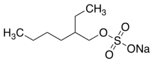
Sodium 2-Ethylhexyl Sulfate (40%-60% in Water)
$88.84 Add to cart View Product DetailsMolecular Formula : C8H17NaO4S
-
Sodium 3-Sulfobenzoate
$163.88 Add to cart View Product DetailsMolecular Formula : C7 H5 O5 S . Na