50g
Showing 201–250 of 320 results
-

KGF/FGF-7, Human
$271.69 Add to cart View Product DetailsKeratinocyte Growth Factor (KGF) is a highly specific epithelial mitogen produced by fibroblasts and mesenchymal stem cells. KGF belongs to the heparin binding Fibroblast Growth Factor (FGF) family, and is known as FGF-7. However, in contrast to the FGF-1, which binds to all known FGF receptors with high affinity, KGF only binds to a splice variant of an FGF receptor, FGFR2-IIIb. FGFR2-IIIb is produced by most of the epithelial cells, indicating that KGF plays roles as a paracrine mediator. KGF induces the differen-tiation and proliferation of various epithelial cells, including keratinocytes in the epidermis, hair follicles and sebaceous glands, and is responsible for the wound repairs of various tissues, including lung, bladder, and kidney.
-
KGF/FGF-7, Mouse
$155.25 Add to cart View Product DetailsKeratinocyte Growth Factor (KGF) is a highly specific epithelial mitogen produced by fibroblasts and mesenchymal stem cells. KGF belongs to the heparin binding Fibroblast Growth Factor (FGF) family, and is known as FGF-7. However, in contrast to FGF-1, which binds to all known FGF receptors with high affinity, KGF only binds to a splice variant of the FGF receptor, FGFR2-IIIb. FGFR2-IIIb is expressedby most epithelial cells, indicating KGF’s roleas a paracrine mediator. KGF induces the differentiation and proliferation of various epithelial cells such as keratinocytes in the epidermis, hair follicles and sebaceous glands., KGF is also responsible for wound repair of various tissuesincluding lung, bladder, and kidney.
-

KLK7, His, Mouse
$189.75 Add to cart View Product DetailsKallikrein-related peptidase 7 (KLK7) is a serine protease and was initially purified from the epidermis and characterised as stratum corneum chymotryptic enzyme (SCCE). It was later identified as the seventh member of the human kallikrein family. KLK7 is secreted as an inactive zymogen in the stratum granulosum layer of the epidermis and may be activated by KLK5 or matriptase. Once active, KLK7 is able to cleave desmocollin and corneodesmosin, indicating a role for KLK7 in maintaining skin homeostasis.
-
KRAS, His, Human (G12C)
$137.14 Add to cart View Product DetailsThe KRAS gene provides instructions for making a protein called K-Ras, part of the RAS/MAPK pathway. The protein relays signals from outside the cell to the cell’s nucleus. These signals instruct the cell to grow and divide (proliferate) or to mature and take on specialized functions (differentiate). The K-Ras protein is a GTPase, which means it converts a molecule called GTP into another molecule called GDP. In this way the K-Ras protein acts like a switch that is turned on and off by the GTP and GDP molecules. KRAS is usually tethered to cell membranes because of the presence of an isoprene group on its C-terminus. There are two protein products of the KRAS gene in mammalian cells that result from the use of alternative exon 4 (exon 4A and 4B respectively): K-Ras4A and K-Ras4B, these proteins have different structure in their C-terminal region and use different mechanisms to localize to cellular membranes including the plasma membrane.
-
KRAS, His, Human (G12D)
$137.14 Add to cart View Product DetailsThe KRAS gene provides instructions for making a protein called K-Ras, part of the RAS/MAPK pathway. The protein relays signals from outside the cell to the cell’s nucleus. These signals instruct the cell to grow and divide (proliferate) or to mature and take on specialized functions (differentiate). The K-Ras protein is a GTPase, which means it converts a molecule called GTP into another molecule called GDP. In this way the K-Ras protein acts like a switch that is turned on and off by the GTP and GDP molecules. KRAS is usually tethered to cell membranes because of the presence of an isoprene group on its C-terminus. There are two protein products of the KRAS gene in mammalian cells that result from the use of alternative exon 4 (exon 4A and 4B respectively): K-Ras4A and K-Ras4B, these proteins have different structure in their C-terminal region and use different mechanisms to localize to cellular membranes including the plasma membrane.
-

L-Leucine
$48.13 Add to cart View Product DetailsL-Leucine
-
Lanthanum Oxide, Powder
$277.17 Add to cart View Product DetailsLanthanum Oxide, Powder
-
LIF, Human
$224.25 Add to cart View Product DetailsLeukemia Inhibitory Factor (LIF) is a pleiotropic cytokine belonging to the long four-helix bundle cytokine superfamily. LIF shares tertiary structure with several other cytokines, including Interleukin-6 (IL-6), Oncostatin M, ciliary neurotropic factor, and cardiotrophin-1, and their functions in vivo are also redundant to some extent. LIF can bind to the common receptor of IL-6 subfamily, gp130, and then recruit its own receptor LIF Receptor to form a ternary complex. The basal expression of LIF in vivo is low; and its expression is induced by pro-inflammatory factors, including lipopolysaccharide, IL-1, and IL-17, and inhibited by anti-inflammatory agents, including IL-4 and IL-13. The functions of LIF include proliferation of primordial germ cells, regulation in blastocyst implantation and early pregnancy, and maintenance of pluripotent embryonic stem cells.
-
LIF, Mouse
$314.81 Add to cart View Product DetailsLeukemia Inhibitory Factor (LIF) is a pleiotropic cytokine belonging to the long four-helix bundle cytokine superfamily. LIF shares tertiary structure with several other cytokines, including Interleukin-6 (IL-6), Oncostatin M, ciliary neurotropic factor, and cardiotrophin-1, and their functions in vivo are also redundant to some extent. LIF can bind to the common receptor of IL-6 subfamily, gp130, and then recruit its own receptor LIF Receptor to form a ternary complex. The basal expression of LIF in vivo is low; and its expression is induced by pro-inflammatory factors, including lipopolysaccharide, IL-1, and IL-17, and inhibited by anti-inflammatory agents, including IL-4 and IL-13. The functions of LIF include proliferation of primordial germ cells, regulation in blastocyst implantation and early pregnancy, and maintenance of pluripotent embryonic stem cells.
-
LIGHT, Human
$137.14 Add to cart View Product DetailsLIGHT, also known as tumor-necrosis factor (TNF) superfamily member 14 (TNFSF14), is predominantly expressed on activated immune cells and some tumor cells. LIGHT (homologous to lymphotoxin, exhibits inducible expression and competes with Herpes Simplex Virus glycoprotein D for Herpes Virus Entry Mediator, a receptor expressed by T cells), is a protein primarily expressed on activated T cells, activated Natural Killer (NK) cells, and immature dendritic cells (DC). LIGHT can function as both a soluble and cell surface-bound type II membrane protein and must be in its homotrimeric form to interact with its two primary functional receptors: Herpes Virus Entry Mediator (HVEM) and Lymphotoxin-β Receptor (LTβR). LIGHT signaling through these receptors have distinct functions that are cell-type dependent, but interactions with both types of receptors have immune-related implications in tumor biology.
-
Lithium Dodecyl Sulfate
$266.20 Add to cart View Product DetailsLithium Dodecyl Sulfate
-
LIX/CXCL5 (74aa), Mouse
$150.94 Add to cart View Product DetailsMouse LIX (C-X-C motif chemokine 5) is a small cytokine belonging to the CXC chemokine family that is cleaved into the following 2 chains [GCP-2(1-78) and GCP-2(9-78)]. Mouse LIX plays a role in reducing sensitivity to sunburn pain in some subjects, and is a potential target which could be used to understand more about pain in other inflammatory conditions. It is most closely related to two highly homologous human neutrophil chemoattractants GCP-2 and ENA-78. The first 78 amino acid residues within the predicted mature mouse LIX shares approximately 61% and 55% amino acid identity with human GCP-2 and ENA-78. This chemokine stimulates the chemotaxis of neutrophils possessing angiogenic properties. It elicits these effects by interacting with the cell surface chemokine receptor CXCR2.
-
M-CSF, Human(CHO-expressed)
$271.69 Add to cart View Product DetailsMacrophage-Colony Stimulating Factor (M-CSF), also known as Colony Stimulating Factor-1 (CSF-1), is a hematopoietic growth factor. It can stimulate the survival, proliferation and differentiation of mononuclear phagocytes, in addition to the spreading and motility of macrophages. In mammals, it exits three isoforms, which invariably share an N-terminal 32-aa signal peptide, a 149-residue growth factor domain, a 21-residue transmembrane region and a 37-aa cytoplasmictail. M-CSF is mainly produced by monocytes, macrophages, fibroblasts, and endothelial cells. M-CSF interaction with its receptor, c-fms, has been implicated in the growth, invasion, and metastasis of of several diseases, including breast and endometrial cancers. The biological activity of human M-CSF is maintained within the 149-aa growth factor domain, and it is only active in the disulfide-linked dimeric form, which is bonded at Cys63.
-

M-CSF, Mouse
$271.69 Add to cart View Product DetailsMacrophage-Colony Stimulating Factor (M-CSF), also known as Colony Stimulating Factor-1 (CSF-1), is a hematopoietic growth factor. It can stimulate the survival, proliferation and differentiation of mononuclear phagocytes, in addition to the spreading and motility of macrophages. In mammals, it exits three isoforms, which invariably share an N-terminal 32-aa signal peptide, a 149-residue growth factor domain, a 21-residue transmembrane region and a 37-aa cytoplasmictail. M-CSF is mainly produced by monocytes, macrophages, fibroblasts, and endothelial cells. M-CSF interaction with its receptor, c-fms, has been implicated in the growth, invasion, and metastasis of of several diseases, including breast and endometrial cancers. The biological activity of human M-CSF is maintained within the 149-aa growth factor domain, and it is only active in the disulfide-linked dimeric form, which is bonded at Cys63.
-
M-CSF, Rat
$271.69 Add to cart View Product DetailsMacrophage-Colony Stimulating Factor (M-CSF), also known as Colony Stimulating Factor-1 (CSF-1), is a hematopoietic growth factor. It can stimulate the survival, proliferation and differentiation of mononuclear phagocytes, in addition to the spreading and motility of macrophages. In mammals, it exits three isoforms, which invariably share an N-terminal 32-aa signal peptide, a 149-residue growth factor domain, a 21-residue transmembrane region and a 37-aa cytoplasmictail. M-CSF is mainly produced by monocytes, macrophages, fibroblasts, and endothelial cells. M-CSF interaction with its receptor, c-fms, has been implicated in the growth, invasion, and metastasis of of several diseases, including breast and endometrial cancers. The biological activity of human M-CSF is maintained within the 149-aa growth factor domain, and it is only active in the disulfide-linked dimeric form, which is bonded at Cys63.
-

MCP‑3/CCL7, Human(CHO-expressed)
$155.25 Add to cart View Product DetailsChemokine (C-C motif) ligand 7 (CCL7) is a small cytokine that was previously called monocyte-specific chemokine 3 (MCP-3). Due to CCL7 possessing two adjacent N-terminal cysteine residues in its mature form, it is classified within the subfamily of chemokines known as CC chemokines. CCL7 specifically attracts monocytes, and regulates macrophage function. It is produced by certain tumor cell lines and by macrophages. This chemokine is located on chromosome 17 in humans, within a large cluster containing many other CC chemokines and is most closely related to CCL2. CCL7 can signal through the CCR1, CCR2 and CCR3 receptors.
-
MES Free Acid
$59.35 Add to cart View Product DetailsMES Free Acid
-
Methyl Alcohol-d4, 99.8 Atom Percent D
$1,035.81 Add to cart View Product DetailsMethyl Alcohol-d4, 99.8 Atom Percent D
-
Methyl Gallate
$110.75 Add to cart View Product DetailsMethyl Gallate
-
Methylphenidate Hydrochloride (CII), USP
$3,280.48 Add to cart View Product DetailsMethylphenidate Hydrochloride (CII), USP
-
MIF, Human
$155.25 Add to cart View Product DetailsMacrophage Migration Inhibitory Factor (MIF) is a pleiotropic cytokine, existing as a homotrimer in vivo. MIF was originally identified as a T cell derived factor responsible for the inhibition of macrophage migration. However, recently MIF has received much more attention because of its possible roles in angiogenesis and cancer development. MIF is over-expressed in various cancers, including pancreatic, breast, colon, brain, prostate, skin, and lung. The intratumoral expression of MIF is strongly correlated with angiogenic growth factor expression, such as the expression of Interleukin 8 (IL-8) and Vascular Endothelial Growth Factor (VEGF), and with risk of recurrence after resection.
-
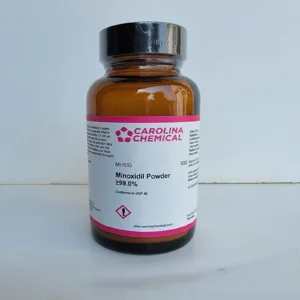
Minoxidil Powder ≥99.0% – 50G
$59.80 Add to cart View Product DetailsCAS Number 38304-91-5 Molecular Formula C9H15N5O Molecular Weight 209.25 -
Mycophenolic Acid
$23,850.90 Add to cart View Product DetailsMycophenolic Acid
-
Nectin-2/CD112 Fc Chimera, Human
$103.50 Add to cart View Product DetailsNectin-2, also known as CD112 (Cluster of Differentiation 226) and PVRL2 (Poliovirus receptor-related 2), is a human plasma membrane glycoprotein. This protein is a modulator of T-cell signaling and can be either a costimulator of T-cell function or a coinhibitor, depending on the receptor it binds to. Upon binding to CD226, it stimulates T-cell proliferation and cytokine production, including that of IL-2, IL-5, IL-10, IL-13, and IFNγ. It can also inhibit T-cell proliferation upon interaction with PVRIG. Nectin-2 also acts as a receptor for herpes simplex virus 1 (HHV-1) mutant Rid1, herpes simplex virus 1 (HHV-2) and pseudorabies virus (PRV).
-
NGF R, Human
$133.69 Add to cart View Product DetailsNGF Receptor, also known as Gp80-LNGFR, p75 ICD, CD271 and TNFRSF16, is a type I transmembrane protein belonging to the TNF receptor family. It is expressed by both neuronal and non-neuronal cells. Signaling through NGF Receptor has been shown to regulate gene expression, cell migration and death. A truncated NGF Receptor containing only the extracellular domain has been detected in plasma, amniotic fluid and urine, and acts as a potent NGF antagonist.
-

Noggin Fc Chimera, Human
$90.56 Add to cart View Product DetailsNoggin, also known as NOG, is a homodimeric glycoprotein that bindsto and modulates the activity of TGF-beta family ligands. It is expressed in condensing cartilage and immature chondrocytes. Noggin antagonizes bone morphogenetic protein (BMP) activities by blocking epitopes on BMPs needed for binding to their receptors. Noggin has been shown to be involved in many developmental processes, such as neural tube formation and joint formation. During development, Noggin diffuses through extracellular matrices and forms morphogenic gradients, regulating cellular responses dependent on the local concentration of the signaling molecule.
-
Noggin, Human(CHO-expressed)
$193.20 Add to cart View Product DetailsNoggin, also known as NOG, is a homodimeric glycoprotein that bindsto and modulates the activity of TGF-beta family ligands. It is expressed in condensing cartilage and immature chondrocytes. Noggin antagonizes bone morphogenetic protein (BMP) activities by blocking epitopes on BMPs needed for binding to their receptors. Noggin has been shown to be involved in many developmental processes, such as neural tube formation and joint formation. During development, Noggin diffuses through extracellular matrices and forms morphogenic gradients, regulating cellular responses dependent on the local concentration of the signaling molecule.
-
NRG-1β2, Human
$155.25 Add to cart View Product DetailsNeuregulin is a signaling protein for ErbB2/ErbB4 receptor heterodimers on the cardiac muscle cells, playing an important role in heart structure and function through inducing ErbB2/ErbB4 receptor phosphorylation and cardiomyocyte differentiation. Research on molecular level discovered that recombinant neuregulin could make disturbed myocardial cell structure into order and strengthen the connection between myocardial cells by intercalated discs re-organization.
-
NT-4, Mouse
$271.69 Add to cart View Product DetailsNeurotrophin-4 (NT-4), also known as NT-5, is a neurotrophic factor structurally related to β-NGF, BDNF, and NT-3. Human NT-4 shares 48 – 52% aa sequence identity with human β-NGF, BDNF, and NT-3. Neurotrophins have six conserved cysteine residues that are involved in the formation of three disulfide bonds. NT-4 is expressed highest levels in prostate, lower levels in thymus, placenta, and skeletal muscle. NT-4 binds and induces receptor dimerization and activation of TrkB. NT-4 can signal through TrkB receptors and promotes the survival of peripheral sensory sympathetic neurons.
-
Octafluorocyclopentene
$956.34 Add to cart View Product DetailsOctafluorocyclopentene
-
Orotic Acid, Anhydrous
$171.06 Add to cart View Product DetailsOrotic Acid, Anhydrous
-
OSM (209aa), Human
$271.69 Add to cart View Product DetailsOncostatin M (OSM) is a multifunctional cytokine, and belongs to Interleukin-6 (IL-6) subfamily, which also includes IL-11, leukemia inhibitory factor (LIF), ciliary neurotropic factor, cardiotrophin-1, and novel neurotropin-1. In vivo, OSM is secreted from activated T cells, monocytes, neutrophils, and endothelial cells. OSM is related to LIF, and shares a receptor with LIF in human. Human OSM can bind to gp130 and recruit OSM Receptor β or LIF Receptor β to form a ternary complex. OSM stimulates the growth of different types of cells, including megakaryocytes, fibroblasts, vascular endothelial cells, and T cells. OSM inhibits the proliferation of several cancer cell lines, such as solid tissue tumor cells, lung cancer cells, melanoma cells, and breast cancer cells.
-
OSM (227aa), Human
$271.69 Add to cart View Product DetailsOncostatin M (OSM) is a multifunctional cytokine, and belongs to Interleukin-6 (IL-6) subfamily, including IL-11, leukemia inhibitory factor (LIF), ciliary neurotropic factor, cardiotrophin-1, and novel neurotropin-1. In vivo, OSM is secreted from activated T cells, monocytes, neutrophils, and endothelial cells. OSM is related to LIF, and share a receptor with LIF in human. Human OSM can bind to gp130 and recruit OSM Receptor β or LIF Receptor β to form a ternary complex. OSM stimulates the growth of different types of cells, including megakaryocytes, fibroblasts, vascular endothelial cells, and T cells. On the other hand, OSM inhibits the proliferation of several cancer cell lines, such as solid tissue tumor cells, lung cancer cells, melanoma cells, and breast cancer cells.
-
OSM, Mouse
$271.69 Add to cart View Product DetailsOncostatin M (OSM) is a multifunctional cytokine, and belongs to Interleukin-6 (IL-6) subfamily, which also includes IL-11, leukemia inhibitory factor (LIF), ciliary neurotropic factor, cardiotrophin-1, and novel neurotropin-1. In vivo, OSM is secreted from activated T cells, monocytes, neutrophils, and endothelial cells. OSM is related to LIF, and shares a receptor with LIF in human. Human OSM can bind to gp130 and recruit OSM Receptor β or LIF Receptor β to form a ternary complex. OSM stimulates the growth of different types of cells, including megakaryocytes, fibroblasts, vascular endothelial cells, and T cells. OSM inhibits the proliferation of several cancer cell lines, such as solid tissue tumor cells, lung cancer cells, melanoma cells, and breast cancer cells.
-
OSM, Mouse(HEK 293-expressed)
$271.69 Add to cart View Product DetailsOncostatin M (OSM) is a multifunctional cytokine, and belongs to Interleukin-6 (IL-6) subfamily, which also includes IL-11, leukemia inhibitory factor (LIF), ciliary neurotropic factor, cardiotrophin-1, and novel neurotropin-1. In vivo, OSM is secreted from activated T cells, monocytes, neutrophils, and endothelial cells. OSM is related to LIF, and shares a receptor with LIF in human. Human OSM can bind to gp130 and recruit OSM Receptor β or LIF Receptor β to form a ternary complex. OSM stimulates the growth of different types of cells, including megakaryocytes, fibroblasts, vascular endothelial cells, and T cells. OSM inhibits the proliferation of several cancer cell lines, such as solid tissue tumor cells, lung cancer cells, melanoma cells, and breast cancer cells.
-

PD-1 Fc Chimera, Human
$103.50 Add to cart View Product DetailsProgrammed cell death protein 1, also known as PD-1 and CD279 (cluster of differentiation 279) or PDCD1, is a protein that in humans is encoded by the PDCD1 gene. PD-1 is a cell surface receptor that belongs to the immunoglobulin superfamily and is expressed on T cells and pro-B cells.PD-1 binds two ligands, PD-L1 and PD-L2. PD-1 and its ligands play an important role in down regulating the immune system by preventing the activation of T-cells, which in turn reduces autoimmunity and promotes self-tolerance. The inhibitory effect of PD-1 is accomplished through a dual mechanism of promoting apoptosis (programmed cell death) in antigen specific T-cells in lymph nodes while simultaneously reducing apoptosis in regulatory T cells (suppressor T cells)
-

PD-1 Fc Chimera, Mouse
$103.50 Add to cart View Product DetailsProgrammed cell death protein 1, also known as PD-1 and CD279 (cluster of differentiation 279) or PDCD1, is a protein that in humans is encoded by the PDCD1 gene. PD-1 is a cell surface receptor that belongs to the immunoglobulin superfamily and is expressed on T cells and pro-B cells.PD-1 binds two ligands, PD-L1 and PD-L2. PD-1 and its ligands play an important role in down regulating the immune system by preventing the activation of T-cells, which in turn reduces autoimmunity and promotes self-tolerance. The inhibitory effect of PD-1 is accomplished through a dual mechanism of promoting apoptosis (programmed cell death) in antigen specific T-cells in lymph nodes while simultaneously reducing apoptosis in regulatory T cells (suppressor T cells).
-
PD-L1 Fc Chimera, Human
$103.50 Add to cart View Product DetailsProgrammed death-ligand 1 (PD-L1) also known as cluster of differentiation 274 (CD274) or B7 homolog 1 (B7-H1), is a protein that in humans is encoded by the CD274 gene. PD-L1 is a 40 kDa type 1 transmembrane protein that has been speculated to play a major role in suppressing the immune system during particular events such as pregnancy, tissue allografts, autoimmune disease and other disease states such as hepatitis. Normally the immune system reacts to foreign antigens where there is some accumulation in the lymph nodes or spleen which triggers a proliferation of antigen-specific CD8+ T cell. The formation of PD-1 receptor / PD-L1 or B7.1 receptor /PD-L1 ligand complex transmits an inhibitory signal which reduces the proliferation of these CD8+ T cells at the lymph nodes and supplementary to that PD-1 is also able to control the accumulation of foreign antigen specific T cells in the lymph nodes through apoptosis which is further mediated by a lower regulation of the gene Bcl-2. PD-L1 binds to its receptor, PD-1, found on activated T cells, B cells, and myeloid cells, to modulate activation or inhibition. Recombinant Human PD-L1(B7-H1) Fc Chimera produced in CHO cells is a polypeptide chain containing 457 amino acids. A fully biologically active molecule, rh PD‑L1(B7-H1) has a molecular mass of 70-72 kDa analyzed by reducing SDS-PAGE and is obtained by chromatographic techniques at GenScript.
-
PD-L1 Fc Chimera, Mouse
$103.50 Add to cart View Product DetailsProgrammed death-ligand 1 (PD-L1) also known as cluster of differentiation 274 (CD274) or B7 homolog 1 (B7-H1), is a protein that in humans is encoded by the CD274 gene. PD-L1 is a 40 kDa type 1 transmembrane protein that has been speculated to play a major role in suppressing the immune system during particular events such as pregnancy, tissue allografts, autoimmune disease and other disease states such as hepatitis. Normally the immune system reacts to foreign antigens where there is some accumulation in the lymph nodes or spleen which triggers a proliferation of antigen-specific CD8+ T cell. The formation of PD-1 receptor / PD-L1 or B7.1 receptor /PD-L1 ligand complex transmits an inhibitory signal which reduces the proliferation of these CD8+ T cells at the lymph nodes and supplementary to that PD-1 is also able to control the accumulation of foreign antigen specific T cells in the lymph nodes through apoptosis which is further mediated by a lower regulation of the gene Bcl-2. PD-L1 binds to its receptor, PD-1, found on activated T cells, B cells, and myeloid cells, to modulate activation or inhibition.
-

PDGF-AA, Mouse
$207.00 Add to cart View Product DetailsPlatelet-Derived Growth Factor-AA (PDGF-AA) is one of five dimers (PDGF-AA, AB, BB, CC, and DD) formed by 4 different PDGF subunits. In chemical terms, platelet-derived growth factor is a dimeric glycoprotein composed of two A (-AA) or two B (-BB) chains or a combination of the two (-AB). The dimeric isoforms PDGFAA, AB and BB are differentially expressed in various cell types, and their effects are mediated through two distinct receptors termed α and β. Differences exist in isoform binding to each receptor. Ingeneral, PDGF isoforms are potent mitogens for connective tissue cells including dermal fibroblasts, glial cells, arterial smooth muscle cells and some epithelial andendothelial cells. In addition to its activity as a mitogen, PDGF is chemotactic for fibroblasts, smooth muscle cells, neutrophils and mononuclear cells. PDGF-AA plays a significant role in blood vessel formation (angiogenesis).
-
PDGF-BB, Human (P. pastoris-expressed)
$258.75 Add to cart View Product DetailsPlatelet-Derived Growth Factor-BB (PDGF-BB) is one of five dimers (PDGF-AA, AB, BB, CC, and DD) formed by 4 different PDGF subunits. In vivo, PDGF-BB is mainly produced in heart and placenta, and predominantly expressed by osteoblasts, fibroblasts, smooth muscle cells, and glial cells. An inactive precursor of PDGF-BB is produced in the endoplasmic reticulum and then activated by a proprotein convertase after secretion. PDGF-BB functions in a paracrine manner and promotes organogenesis, human skeletal development, and wound healing. PDGF-BB also promotes angiogenesis, particularly in the presence of Fibroblast Growth Factor basic. Therefore, PDGF-BB and its related pathways are potential pharmacological targets.
-
PDGF-BB, Mouse
$194.06 Add to cart View Product DetailsPlatelet-Derived Growth Factor-BB (PDGF-BB) is one of five dimers (PDGF-AA, AB, BB, CC, and DD) formed by 4 different PDGF subunits. In vivo, PDGF-BB is mainly produced in heart and placenta, and predominantly expressed by osteoblasts, fibroblasts, smooth muscle cells, and glial cells. An inactive precursor of PDGF-BB is produced in the endoplasmic reticulum and then activated by a proprotein convertase after secretion. PDGF-BB functions in a paracrine manner and promotes organogenesis, human skeletal development, and wound healing. PDGF-BB also promotes angiogenesis, particularly in the presence of Fibroblast Growth Factor basic. Therefore, PDGF-BB and its related pathways are potential pharmacological targets.
-
PDGF-BB, Rat
$194.06 Add to cart View Product DetailsPlatelet-Derived Growth Factor-BB (PDGF-BB) is one of five dimers (PDGF-AA, AB, BB, CC, and DD) formed by 4 different PDGF subunits. In vivo, PDGF-BB is mainly produced in heart and placenta, and predominantly expressed by osteoblasts, fibroblasts, smooth muscle cells, and glial cells. An inactive precursor of PDGF-BB is produced in the endoplasmic reticulum and then activated by a proprotein convertase after secretion. PDGF-BB functions in a paracrine manner and promotes organogenesis, human skeletal development, and wound healing. PDGF-BB also promotes angiogenesis, particularly in the presence of Fibroblast Growth Factor basic. Therefore, PDGF-BB and its related pathways are potential pharmacological targets.
-
PDGF-CC, Human
$155.25 Add to cart View Product DetailsPlatelet-Derived Growth Factor (PDGF) is a potent mitogen for a wide range of cell types including fibroblasts, smooth muscle, connective tissue, bone and cartilage cells, and some blood cells. The PDGF is involved in a number of biological processes, including hyperplasia, chemotaxis, embryonic neuron development, and respiratory tubule epithelial cell development. The PDGF family consists of proteins derived from four genes (PDGF -A, -B, -C, and -D) that form four disulfide-linked homodimers (PDGF-AA, -BB, -CC, and -DD) and one heterodimer (PDGF-AB).
-
PDGF-DD, Human
$194.06 Add to cart View Product DetailsPDGF-DD, also known as platelet-derived growth factor D, IEGF and SCDGFB, is asecreted growth factor belonging to the PDGF/VEGFfamily. It is highly expressed in the heart, pancreas, adrenal glands and ovary. PDGF-DD forms functional homodimers that bind and induce PDGF Rβ homodimers and PDGF Rα/β heterodimers that promote intracellular signaling. This plays an important role in the regulation of cell differentiation, migration and survival. It has also been reported that PDGF-DD can induce monocyte and macrophage recruitment, increase interstitial pressure and facilitate wound healing.
-
PF-4/CXCL4, Human
$155.25 Add to cart View Product DetailsPlatelet factor 4, also known as CXCL4, is expressed in megakaryocytes and stored in the α-granules of platelets. Recombinant human PF-4 is a 7.8 kDa protein containing 70 amino acid residues, including the four highly conserved residues present in CXC chemokines. Platelet factor 4 can be antiproliferative and antiangiogenic, at least in part via interfering with FGF2 and VEGF heparin binding and thus inhibiting their signaling. However, it can also be proinflammatory and proatherogenic through multiple effects on monocytes, macrophages and endothelial cells.
-

Phenylpiracetam Hydrazide Powder – 50G
$170.20 Add to cart View Product DetailsCAS Number 77472-71-0 Molecular Weight 233.27 Molecular Formula C12H15N3O2 -
Phosphorus Oxychloride, High Purity
$2,630.02 Add to cart View Product DetailsPhosphorus Oxychloride, High Purity
-
Phytochol, cGMP Manufactured
$7,205.51 Add to cart View Product DetailsPhytochol, cGMP Manufactured