2
Showing 751–800 of 1694 results
-

eBlot L1 Transfer Cassette
$625.31 Add to cart View Product DetailsTransfer Cassette
-

Economy Class B Double Metric Scale Cylinders, 1000 ml
$117.73 Add to cart View Product DetailsEconomy Class B Double Metric Scale Cylinders, 1000 ml
-
Economy Class B Double Metric Scale Cylinders, 1000 ml
$470.93 Add to cart View Product DetailsEconomy Class B Double Metric Scale Cylinders, 1000 ml
-
Economy Class B Double Metric Scale Cylinders, 500 ml
$69.00 Add to cart View Product DetailsEconomy Class B Double Metric Scale Cylinders, 500 ml
-
Economy Class B Double Metric Scale Cylinders, 500 ml
$413.91 Add to cart View Product DetailsEconomy Class B Double Metric Scale Cylinders, 500 ml
-
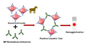
Equine Anti-Globulin Test
$227.06 Add to cart View Product DetailsThe anti-globulin or Coombs’ test is widely used to detect the presence of antibody in the diagnosis of autoimmune hemolytic anemia.
-
Erlenmeyer Flasks, Conical, Narrow Mouth, Borosilicate, ISO 1773, 5000 mL
$176.42 Add to cart View Product DetailsErlenmeyer Flasks, Conical, Narrow Mouth, Borosilicate, ISO 1773, 5000 mL
-
eStain Gel Holder
$327.75 Add to cart View Product DetailseStain Gel Holder for eStain L1, eStain L1c, and eStain LG.
-
Extractor EtBr System, starter pack, 2/pk
$66.30 Add to cart View Product DetailsExtractor EtBr System, starter pack, 2/pk
-

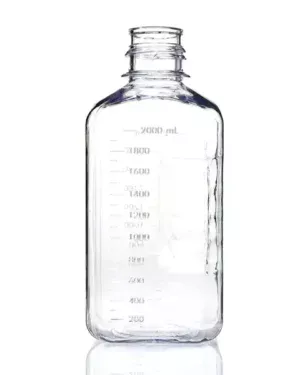
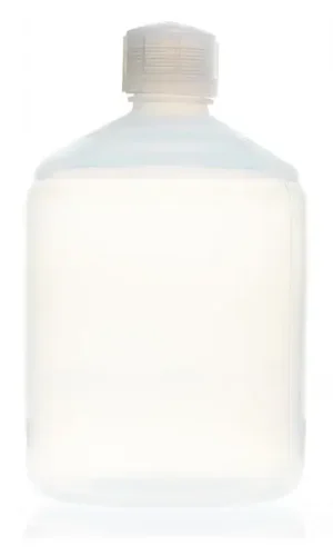
EZBio® 2 L (2,000 mL) PC Media Bottles, Non-Sterile, Autoclavable Square Storage Bottles with 53 mm (53B) Neck, Without Caps, 6/PK
$138.76 Add to cart View Product DetailsEZBio® 2 L (2,000 mL) PC Media Bottles, Non-Sterile, Autoclavable Square Storage Bottles with 53 mm (53B) Neck, Without Caps, 6/PK
-

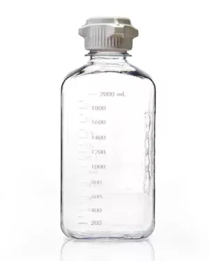
EZBio® 2 L (2,000 mL) PC Media Bottles, Sterile, Autoclavable Square Storage Bottles with 53 mm (53B) White Closed VersaCap Closures, 6/PK
$190.33 Add to cart View Product DetailsEZBio® 2 L (2,000 mL) PC Media Bottles, Sterile, Autoclavable Square Storage Bottles with 53 mm (53B) White Closed VersaCap Closures, 6/PK
-
EZBio® 2 L (2,000 mL) PETG Media Bottles, Non-Sterile, Square Storage Bottles with 53 mm (53B) Neck, Without Caps, 6/PK
$134.19 Add to cart View Product DetailsEZBio® 2 L (2,000 mL) PETG Media Bottles, Non-Sterile, Square Storage Bottles with 53 mm (53B) Neck, Without Caps, 6/PK
-
EZBio® 2 L (2,000 mL) PETG Media Bottles, Sterile, Square Storage Bottles with 53 mm (53B) White Closed VersaCap Closures, 6/PK
$177.18 Add to cart View Product DetailsEZBio® 2 L (2,000 mL) PETG Media Bottles, Sterile, Square Storage Bottles with 53 mm (53B) White Closed VersaCap Closures, 6/PK
-
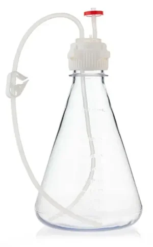
EZBio® Flask Assembly 2L VersaCap® 53B, PC, Baffle EZBio Tubing w/ Plug and Vented w/ Diptube
$971.05 Add to cart View Product DetailsEZBio® Flask Assembly 2L VersaCap® 53B, PC, Baffle EZBio Tubing w/ Plug and Vented w/ Diptube
-

EZBio® Flask Assembly 2L VersaCap® 53B, PC, No Baffle EZBio Tubing w/ Plug and Vented w/ Diptube
$971.05 Add to cart View Product DetailsEZBio® Flask Assembly 2L VersaCap® 53B, PC, No Baffle EZBio Tubing w/ Plug and Vented w/ Diptube
-
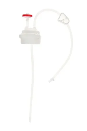
EZBio® Flask Cap Assembly 2L VersaCap® 53B, PC, EZBio Tubing w/ Plug and Vented w/ Diptube
$1,428.99 Add to cart View Product DetailsEZBio® Flask Cap Assembly 2L VersaCap® 53B, PC, EZBio Tubing w/ Plug and Vented w/ Diptube
-
EZBio® Pure PFA Bottle, 2 L (2,000 mL) Low Temperature Resistant, Autoclavable ultraclean Bottle with GL45 (45 mm) Cap, 1/EA
$474.33 Add to cart View Product DetailsEZBio® Pure PFA Bottle, 2 L (2,000 mL) Low Temperature Resistant, Autoclavable ultraclean Bottle with GL45 (45 mm) Cap, 1/EA
-

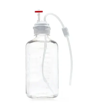
EZBio® Single Use Media Bottle Assembly, 2000 mL (2 L) Sterilized PETG Bottles for Aseptic Sampling and Sterile Storage, 53 mm (53B) VersaCap with Vent and Dip Tube, 10/CS
$1,018.39 Add to cart View Product DetailsEZBio® Single Use Media Bottle Assembly, 2000 mL (2 L) Sterilized PETG Bottles for Aseptic Sampling and Sterile Storage, 53 mm (53B) VersaCap with Vent and Dip Tube, 10/CS
-
EZBio® Single Use Media Bottle Assembly, 2000 mL (2 L) Sterilized Polycarbonate (PC) Bottles for Aseptic Sampling and Sterile Storage, 53 mm (53B) VersaCap with Vent and Dip Tube, 10/CS
$1,022.88 Add to cart View Product DetailsEZBio® Single Use Media Bottle Assembly, 2000 mL (2 L) Sterilized Polycarbonate (PC) Bottles for Aseptic Sampling and Sterile Storage, 53 mm (53B) VersaCap with Vent and Dip Tube, 10/CS
-
EZwaste® HPLC Solvent Waste System, Breakthrough Indicator for Detecting Carbon Filter Saturation, 2/PK
$115.20 Add to cart View Product DetailsEZwaste® HPLC Solvent Waste System, Breakthrough Indicator for Detecting Carbon Filter Saturation, 2/PK
-
EZwaste® HPLC Solvent Waste System, Large Size Replacement Chemical Exhaust Filter, Activated Carbon, 2/PK
$440.61 Add to cart View Product DetailsEZwaste® HPLC Solvent Waste System, Large Size Replacement Chemical Exhaust Filter, Activated Carbon, 2/PK
-
EZwaste® HPLC Solvent Waste System, Standard Size Replacement Chemical Exhaust Filter, Activated Carbon, 2/PK
$148.89 Add to cart View Product DetailsEZwaste® HPLC Solvent Waste System, Standard Size Replacement Chemical Exhaust Filter, Activated Carbon, 2/PK
-
EZwaste® HPLC Solvent Waste System, XLarge Size Replacement Chemical Exhaust Filter, Activated Carbon, 2/PK
$670.77 Add to cart View Product DetailsEZwaste® HPLC Solvent Waste System, XLarge Size Replacement Chemical Exhaust Filter, Activated Carbon, 2/PK
-

F(ab’)2 Goat Anti-Human Kappa Chain (bound)
$326.51 Add to cart View Product DetailsLyophilized goat F(ab’)2 fragment to human kappa chain (bound) and buffer salts.
-

Falcon 525cm2 Rectangular Straight Neck Cell Culture Multi-Flask, 3-layer with Vented Cap
$289.92 Add to cart View Product DetailsFalcon 525cm2 Rectangular Straight Neck Cell Culture Multi-Flask, 3-layer with Vented Cap
-
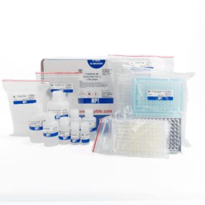
FastDNA-96 Fecal DNA extraction Kit
$1,074.04 Add to cart View Product DetailsDesigned to isolate PCR-ready genomic DNA from fecal and soil samples for a quick and efficient purification of host and microbial DNA free of inhibitors.
-
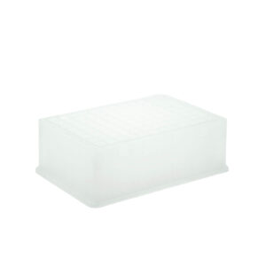
FastPrep 96 PCR plate holder for DNA shearing
$40.97 Add to cart View Product DetailsFastPrep 96 PCR plate holder for DNA shearing
-
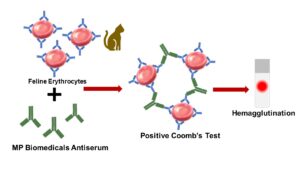
Feline Anti-Globulin Test
$255.15 Add to cart View Product DetailsThe anti-globulin or Coombs’ test is widely used to detect the presence of antibody in the diagnosis of autoimmune hemolytic anemia.
-
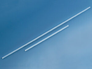
Filling tube for seripettor, 250mm, pack of 2
$12.72 Add to cart View Product DetailsFilling tube for seripettor, 250mm, pack of 2
-
Filling tube for seripettor, 500mm, pack of 2
$18.03 Add to cart View Product DetailsFilling tube for seripettor, 500mm, pack of 2
-
FITC Affinity Purified F(ab’)2 Goat Anti-Mouse IgG, IgA, IgM
$372.69 Add to cart View Product DetailsFluorescein-5-isothiocyanate (FITC “Isomer I”)-conjugated goat affinity purified F(ab’)2 fragments to mouse immunoglobulins (IgG,IgA,IgM) and buffer salts.
-
FITC Affinity Purified Goat Anti-Mouse IgG (H&L)
$198.82 Add to cart View Product DetailsLyophilized fluorescein-5-isothiocyanate (FITC “Isomer I”)-conjugated goat affinity purified antibody to mouse IgG (whole molecule) and buffer salts.
-
FITC Affinity Purified Goat Anti-Mouse IgM (m specific)
$557.68 Add to cart View Product DetailsFITC Conjugated, affinity purified, goat antibody to mouse IgM (µ specific)
-
FITC Affinity Purified Goat Anti-Mouse Immunoglobulins
$766.12 Add to cart View Product DetailsFluorescein-5-isothiocyanate (FITC “Isomer I”)-conjugated goat affinity purified antibody to mouse immunoglobulins (IgG,IgA,IgM) and buffer salts.
-
FITC Conjugated Goat IgG Fraction to Human IgG (Whole Molecu
$195.90 Add to cart View Product DetailsLyophilized fluorescein-5-isothiocyanate (FITC “Isomer I”)-conjugated goat IgG fraction to human IgG (whole molecule) and buffer salts.
-
FITC F(ab’)2 Goat Anti-Human Complement C3
$402.60 Add to cart View Product DetailsLyophilized fluorescein-5-isothiocyanate (FITC “Isomer I”)-conjugated goat F(ab’)2 fragment to human complement C3 and buffer salts.
-
FITC F(ab’)2 Goat Anti-Human Lambda Chain(Bound)
$545.10 Add to cart View Product DetailsLyophilized fluorescein-5-isothiocyanate (FITC “Isomer I”)-conjugated goat F(ab’)2 fragment to human lambda chain (bound) and buffer salts.
-
FITC F(ab’)2 Goat Anti-Mouse Complement C3
$1,010.42 Add to cart View Product DetailsLyophilized fluorescein-5-isothiocyanate (FITC “Isomer I”)-conjugated goat F(ab’)2 fragment to mouse complement C3 and buffer salts.
-
FITC F(ab’)2 Rabbit Anti-Mouse IgG (H&L)
$372.60 Add to cart View Product DetailsLyophilized fluorescein-5-isothiocyanate (FITC “Isomer I”)-conjugated rabbit F(ab’)2 fragment to mouse IgG (whole molecule) and buffer salts
-
FITC Goat Affinity Purified Antibody to Dog IgG (whole molec
$249.06 Add to cart View Product DetailsAffinity-purified goat antibody to dog IgG (whole molecule)
-
FITC Goat F(ab’)2 Fragment to Mouse IgM (µ Chain)
$190.18 Add to cart View Product DetailsLyophilized goat antibody to mouse IgM (Mu specific), FITC conjugated
-
FITC IgG Goat Anti-Cat IgG (H&L)
$244.31 Add to cart View Product DetailsLyophilized fluorescein-5-isothiocyanate (FITC “Isomer I”)-conjugated goat IgG fraction to cat IgG (whole molecule) and buffer salts
-
FITC IgG Goat Anti-Dog IgM (µ CHAIN)
$187.85 Add to cart View Product DetailsLyophilized fluorescein-5-isothiocyanate (FITC “Isomer I”)-conjugated goat IgG fraction to dog IgM (µ chain) and buffer salts.
-
FITC IgG Goat Anti-Guinea Pig Complement C3
$623.09 Add to cart View Product DetailsLyophilized fluorescein-5-isothiocyanate (FITC “Isomer I”)-conjugated goat IgG fraction to guinea pig complement C3 and buffer salts.
-
FITC IgG Goat Anti-Human Complement C1Q
$410.44 Add to cart View Product DetailsLyophilized fluorescein-5-isothiocyanate (FITC “Isomer I”)-conjugated goat IgG fraction to human complement C1q and buffer salts.
-
FITC IgG Goat Anti-Human Complement C3
$344.78 Add to cart View Product DetailsLyophilized fluorescein-5-isothiocyanate (FITC “Isomer I”)-conjugated goat IgG fraction to human complement C3 and buffer salts.
-
FITC IgG Goat Anti-Human Complement C4
$188.37 Add to cart View Product DetailsLyophilized fluorescein-5-isothiocyanate (FITC “Isomer I”)-conjugated goat IgG fraction to human complement C4 and buffer salts.
-
FITC IgG Goat Anti-Human Fibrinogen
$292.46 Add to cart View Product DetailsLyophilized fluorescein-5-isothiocyanate (FITC “Isomer I”)-conjugated goat IgG fraction to human fibrinogen and buffer salts.
-
FITC IgG Goat Anti-Human IgA(α chain)
$370.73 Add to cart View Product DetailsLyophilized fluorescein-5-isothiocyanate (FITC “Isomer I”)-conjugated goat IgG fraction to human IgA (α chain) and buffer salts.
-
FITC IgG Goat Anti-Human IgG F(ab’)2
$196.33 Add to cart View Product DetailsLyophilized fluorescein-5-isothiocyanate (FITC “Isomer I”)-conjugated goat IgG fraction to human IgG F(ab’)2 and buffer salts.