1 each
Showing 101–150 of 294 results
-
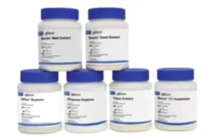
Gibco™Starter Pak No. 1
$379.14 Add to cart View Product DetailsStarter Pak No. 1 features a combination of yeast- and soy-based peptones. Three of the products in this pack have been ultra-filtered to reduce endotoxin levels. The yeast products add a mixture of peptides, amino acids, carbohydrates (simple and complex), nucleosides, and vitamins to any medium formulation. Starter Pak No. 1 provides conveniently packaged, 100-g, non-GMP samples of these peptones that are best suited for use in human and animal health applications and contains:
• Difco TC Yeastolate UF and Bacto TC Yeastolate—these peptones are ideal for CHO-based applications of biotherapeutic monoclonal antibodies and recombinant proteins
• Difco Yeast Extract UF and Bacto Yeast Extract, Technical—these peptones support optimal growth of many microbial species for a variety of human and animal health vaccines
• Difco Phytone Supplement, UF—this enzymatic digest of soy is an excellent, nutritious source of carbohydrates and is used in mammalian cell culture. This peptone works well alone and when blended with yeast-based peptones.
Our Starter Paks provide conveniently packaged samples of the peptones most commonly used in mammalian cell culture and microbial fermentation bioproduction processes. Starter Paks are tailored for specific applications, including the production of monoclonal antibodies, recombinant proteins, and vaccines. -
Gibco™Starter Pak No. 2
$445.54 Add to cart View Product DetailsStarter Pak No. 2 contain a variety of animal origin and animal origin-free peptones that offer many essential nutrients needed for the production of human and animal vaccines. It provides conveniently packaged, 100-g, non-GMP samples of these peptones that are best suited for vaccine production and contains:
• Bacto Yeast Extract—this yeast has the highest level of carbohydrates of our yeast products and works well in a variety of human and animal health vaccines
• Phytone Peptone and Difco Soytone—both enzymatic digests of soy, these peptones also are a nutritious source of carbohydrates. These products work well in microbial fermentation processes as well as in mammalian cell culture processes such as CHO. Blending these soy peptones with yeast peptones has been shown to provide additional benefit to cultures.
• Bacto Proteose Peptone No. 2 and Bacto Proteose Peptone No 3—these enzymatic digests of porcine protein provide nutrition for fastidious microorganisms. Bacto Proteose Peptone No. 3 can replace serum in many applications and helps increase monoclonal antibody and recombinant protein production in CHO cells.
• Bacto Casamino Acids—this supplement has low salt and iron content, making it an excellent supplement for media formulations for which nitrogen requirements are minimal -

Gibco™Starter Pak No. 3
$379.16 Add to cart View Product DetailsStarter Pak No. 3 provides a variety of yeast and soy products, ideal for processes where an animal origin-free medium is preferred. It provides conveniently packaged, 100-g, non-GMP samples of these peptones that are best suited for human and animal vaccine production and contains:
• Bacto Yeast Extract and Gibco Yeast Extract—these peptones contain a mixture of peptides, amino acids, carbohydrates, and vitamins to support optimal growth in microbial species, and are ideal for the production of vaccines
• Bacto TC Yeastolate—this peptone is ideal for CHO-based applications of biotherapeutic monoclonal antibodies and recombinant proteins, as well as vaccine applications
• Phytone Peptone and Difco Soytone—these soy-based supplements provide a nutritious source of carbohydrates for successful use in microbial fermentation. Blending these soy peptones with yeast peptones has been shown to provide additional benefit to cultures.
• Bacto Malt Extract—a water-soluble portion of malted barley, this peptone also provides carbohydrates for a variety of microbial fermentation processes -
Hand Pump for 1 Gallon 7× Bottles
$7.06 Add to cart View Product DetailsDispenses 1 fl. oz
-

HBSS, no calcium, no magnesium, no phenol red
$308.20 Add to cart View Product DetailsHank’s Balanced Salt Solution (HBSS) formulations without calcium and magnesium are required for rinsing chelators from the culture before cell dissociation. This HBSS is modified as follows: with glucose; without calcium magnesium, or phenol. Animal origin-free. The complete formulation is available.
-
- Sustainable packaging
n
Cell Culture, Mammalian Cell Culture
Order Info
Shipping Condition: Room Temperature
-
-

HiPrep adapter for 48 x 2 mL tube holder on FastPrep-24
$2,020.84 Add to cart View Product DetailsAdapter for FastPrep-24 instrument. Holds 48 x 2 mL tubes for room temperature cell grinding and lysis
1,757.25 -

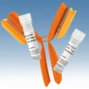
HLA-A2 Monoclonal Antibody (BB7.2), PE
$484.15 Add to cart View Product DetailsThis antibody recognizes an epitope at the C-terminus of alpha-2 helix and a turn on one of the underlying beta strands within the human HLA-A2 histocompatibility antigen.
nHLA-A2 (44 kDa) is the most frequent HLA-A allele in human ethnic populations. HLA-A, together with HLA-B and HLA-C, represent human HLA class I major histocompatibility (MHC) antigens. These intrinsic membrane glycoproteins are expressed on nucleated cells and noncovalently associate with an invariant beta2 microglobulin. They carry foreign determits important for immune recognition by cytotoxic T cells, thus important for anti-viral and anti-tumour defence.
-
Human ACTB (Beta Actin) Endogenous Control (FAM™/MGB probe, non-primer limited)
$449.39 Add to cart View Product DetailsThe Applied Biosystems™ Human ACTB (actin, beta) Endogenous Control (FAM™ Dye / MGB Probe, Non-Primer Limited) is intended as an endogenous control. It allows relative gene expression quantification in cDNA samples when used with other TaqMan™ Gene Expression Assays. Probe is labeled with 6FAM™ dye – MGB and the primers are not limited. Can be used for singleplex PCR reactions. Endogenous control is to be used with Inventoried and Non-Inventoried TaqMan™ Gene Expression Assays, Custom TaqMan™ Gene Expression Assays, and/or Custom TaqMan™ Primers and Probes.
Assay Details:
Gene Symbol: ACTB
RefSeq: NM_001101.2
Probe Exon Location:1
Amplicon Size: 171
Corresponding TaqMan Assay ID: Hs99999903_m1
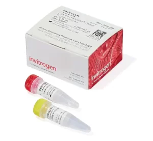
TaqMan™ Endogenous Controls
Eliminate months of assay design, formulation, and testing by using TaqMan™ Endogenous Controls as your controls to quantify gene expression. This convenient collection of pre-designed probe and primer sets enables you to normalize the amount of sample RNA or DNA in a reaction.
Complete Solution for Quantitative Gene Expression
Having a hard time deciding what controls to use to quantify gene expression — even with detailed information on biological systems? Now, with TaqMan™ Endogenous Controls, you can avoid all the trial-and-error of selecting controls for most common human, mouse, rat, and eukaryotic genes.
Simple to Use
All components of the TaqMan™ Endogenous Controls are QC tested, formulated into a single 20X mix, and functionally tested. The controls are not only simple to use, but they are also fully compatible with universal conditions for two-step RT-PCR. Just add TaqMan™ Universal PCR Master Mix (with or without AmpErase™ UNG) and your cDNA sample to generate sensitive, reproducible, and truly quantitative gene expression data on Applied Biosystems instruments including the Applied Biosystems 7900HT, 7300, 7500 Real-Time PCR Systems, and the 7000 and 7700 Sequence Detection Systems.
Flexible Offering
We build each endogenous control using our proven 5′ nuclease chemistry. For maximum flexibility, you can choose between two different reporter dyes and two quenchers:-
- A FAM™ dye-labeled TaqMan™ MGB probe (250nM, final concentration) and two unlabeled PCR primers (900nM each)
n
-
- A VIC™ dye-labeled TaqMan™ MGB probe (250nM, final concentration) and two unlabeled PCR primers (150nM each y primer limited)
n
-
- A VIC™ dye-labeled TAMRA™ probe (250nM, final concentration) and two unlabeled PCR primers (150nM each y primer limited)
Choosing the Right Endogenous Control
Endogenous controls can normalize the expression levels of target genes by correcting differences in the amount of cDNA that is loaded into PCR reaction wells. For best results, verify that the endogenous control is consistently expressed in the sample set to be tested. Endogenous control expression must be uniform across all samples in the study. For multiplexing, ensure that the gene expression level of the endogenous control is greater than that of the target.
Multiplex vs. Singleplex PCR
All TaqMan™ Endogenous Controls that contain probes labeled with the VIC™ reporter dye are primer limited. This allows multiplexing of TaqMan™ Endogenous Controls with target gene expression assays, provided that the control gene is more abundantly expressed than the target gene. All TaqMan™ Endogenous Controls that contain probes labeled with the FAM™ reporter dye are not primer limited and are not intended for multiplexing .
For Research Use Only. Not for use in diagnostic procedures.
- A VIC™ dye-labeled TAMRA™ probe (250nM, final concentration) and two unlabeled PCR primers (150nM each y primer limited)
n
-
-

IgG (H+L) Horse anti-Mouse, Biotin, Invitrogen™ SKU: 31806
$292.10 Add to cart View Product DetailsProduct No. 31806 has been successfully used in Western blot, IF, ICC, IHC and ISH applications. Product No. 31806 reacts with the heavy chains of mouse IgG and with the light chains common to most mouse immunoglobulins. This antibody cross-reacts with mouse IgM (50%) hamster IgG, rat IgG (12%) rat IgM (6%) dog IgG, guinea pig IgG, swine IgG (3%) Rhesus monkey IgG (1%). It has minimal cross-reactivity with immunoglobulins from other species. Store product at 4°C until opened. To extend the shelf-life of this product, add an equal volume of glycerol to make a final concentration of approximately 50% glycerol and store at -20°C.
nThermo Scientific Anti-Mouse secondary antibodies are affinity-purified antibodies with well-characterized specificity for mouse immunoglobulins and are useful in the detection, sorting or purification of its specified target. Secondary antibodies offer increased versatility enabling users to use many detection systems (e.g. HRP, AP, fluorescence). They can also provide greater sensitivity through signal amplification as multiple secondary antibodies can bind to a single primary antibody. Most commonly, secondary antibodies are generated by immunizing the host animal with a pooled population of immunoglobulins from the target species and can be further purified and modified (i.e. immunoaffinity chromatography, antibody fragmentation, label conjugation, etc.) to generate highly specific reagents.
-
IgG (H+L) Horse anti-Mouse, Invitrogen™ SKU: 31181
$285.20 Add to cart View Product DetailsProduct No. 31181 has been successfully used in Western blot, IF, ICC, IHC, IP and ISH applications. Product No. 31181 reacts with heavy chains of mouse IgG and with the light chains common to most mouse immunoglobulins. Based on solid-phase enzyme immunoassay, this antibody cross-reacts with rat IgG and mouse IgM (25%), hamster IgG (12%), guinea pig IgG and rat IgM (6%), bovine IgM, dog IgM, goat IgM, human IgM, Rhesus monkey IgG, sheep IgG, sheep IgM, swine IgG (1%). It has minimum cross-reactivity with immunoglobulins from other species. The activity measured by a solid phase binding assay is 94%. Store lyophilized product at 4°C until opened. To extend the shelf-life of this product, freeze undiluted product in small aliquots. Avoid repeated freezing and thawing.
nThermo Scientific Anti-Mouse secondary antibodies are affinity-purified antibodies with well-characterized specificity for mouse immunoglobulins and are useful in the detection, sorting or purification of its specified target. Secondary antibodies offer increased versatility enabling users to use many detection systems (e.g. HRP, AP, fluorescence). They can also provide greater sensitivity through signal amplification as multiple secondary antibodies can bind to a single primary antibody. Most commonly, secondary antibodies are generated by immunizing the host animal with a pooled population of immunoglobulins from the target species and can be further purified and modified (i.e. immunoaffinity chromatography, antibody fragmentation, label conjugation, etc.) to generate highly specific reagents.
-

Inlet Tubing 50-250uL Dispenser
$23.77 Add to cart View Product DetailsInlet Tubing 50-250uL Dispenser
-
Invitrogen TrueGuide sgRNA Positive Control, AAVS1 (human), SKU: A35522
$299.28 Add to cart View Product DetailsProvides complete collection of validated positive controls for the TrueGuide Synthetic gRNA product line to help maximize editing performance. This positive control targeting AAVS1 targets intronic region of the AAVS1 locus and has achieved editing efficiencies of up to 60% in variety of human cell lines. AAVS1 controls easily determine the optimal conditions for delivering CRISPR/Cas9 tools to cell type, allowing to move forward in editing experiments and minimize downstream effort to isolate and validate your edited clones.
-

InvitrogenFGF1 Polyclonal Antibody, Biotin, PeproTech®
$449.85 Add to cart View Product DetailsnnAA Sequence of recombinant protein: MFNLPPGNYK KPKLLYCSNG GHFLRILPDG TVDGTRDRSD QHIQLQLSAE SVGEVYIKST ETGQYLAMDT DGLLYGSQTP NEECLFLERL EENHYNTYIS KKHAEKNWFV GLKKNGSCKR GPRTHYGQKA ILFLPLPVSS D.
Preparation: Produced from sera of rabbits immunized with highly pure Recombinant Human FGF-acidic. Anti-Human FGF-acidic-specific antibody was purified by affinity chromatography and then biotinylated.
Sandwich ELISA: To detect hFGF-acidic by sandwich ELISA (using 100 µL/well antibody solution) a concentration of 0.25-1.0 µg/mL of this antibody is required. This biotinylated polyclonal antibody, in conjunction with PeproTech Polyclonal Anti-Human FGF-acidic (500-P17) as a capture antibody, allows the detection of at least 0.2-0.4 ng/well of Recombinant hFGF-acidic.
Western Blot: To detect Human FGF-acidic by Western Blot analysis this antibody can be used at a concentration of 0.1-0.2 µg/mL. Used in conjunction with compatible secondary reagents the detection limit for Recombinant Human FGF-acidic is 1.5-3.0 ng/lane, under either reducing or non-reducing conditions.Target Information
FGF1 is a member of the fibroblast growth factor (FGF) family. FGF family members possess broad mitogenic and cell survival activities, and are involved in a variety of biological processes, including embryonic development, cell growth, morphogenesis, tissue repair, tumor growth and invasion. This protein functions as a modifier of endothelial cell migration and proliferation, as well as an angiogenic factor. It acts as a mitogen for a variety of mesoderm- and neuroectoderm-derived cells in vitro, thus is thought to be involved in organogenesis.
-
InvitrogenFGF1 Polyclonal Antibody, PeproTech®
$351.69 Add to cart View Product DetailsAA Sequence of recombinant protein: MFNLPPGNYK KPKLLYCSNG GHFLRILPDG TVDGTRDRSD QHIQLQLSAE SVGEVYIKST ETGQYLAMDT DGLLYGSQTP NEECLFLERL EENHYNTYIS KKHAEKNWFV GLKKNGSCKR GPRTHYGQKA ILFLPLPVSS D.
Preparation: Produced from sera of rabbits immunized with highly pure Recombinant Human FGF-acidic. Anti-Human FGF-acidic-specific antibody was purified by affinity chromatography employing an immobilized Human FGF-acidic matrix.
Sandwich ELISA: To detect Human FGF-acidic by sandwich ELISA (using 100 µL/well antibody solution) a concentration of 0.5-2.0 µg/mL of this antibody is required. This antigen affinity purified antibody, in conjunction with PeproTech Biotinylated Anti-Human FGF-acidic (500-P17Bt) as a detection antibody, allows the detection of at least 0.2-0.4 ng/well of Recombinant Human FGF-acidic.
Western Blot: To detect Human FGF-acidic by Western Blot analysis this antibody can be used at a concentration of 0.1-0.2 µg/mL. When used in conjunction with compatible secondary reagents, the detection limit for Recombinant Human FGF-acidic is 1.5-3.0 ng/lane, under either reducing or non-reducing conditions. -
Invitrogen™ BODIPY™ TR Ceramide SKU: d7540
$401.35 Add to cart View Product DetailsBeschreibung
BODIPY TR ceramide can be used to stain Golgi apparatus in live cells.
Cell Analysis, Cell Structure, Golgi
Bestellinformation
Shipping Condition: Room Temperature
-
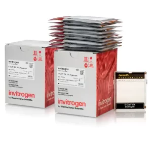
Invitrogen™ E-Gel™ EX Agarose Gels, 2%
$397.90 Add to cart View Product Details-
- Contain a proprietary fluorescent nucleic acid stain (excitation: 490nm, emission: 520nm) that can be viewed with blue light, which minimizes DNA damage and allows detection down to 1ng/band of DNA
n
-
- Complete separation (by molecular weight) in 10 minutes
n
-
- Live monitoring of DNA migration without the hazards of UV light
n
-
- Easy access to the gel for extraction of DNA bands or transfer to membranes for Southern blotting
n
-
- Demonstrate over five-fold greater sensitivity than comparable gels containing ethidium bromide allows less sample use per run
n
-
- Fast analysis of RNA samples to check on their integrity before proceeding with downstream application
n
-
- The gel cassette is designed to open with a Gel Knife, so that the gel inside can be accessed readily for excision of specific bands or transferred to a membrane for Southern blot analysis
n
-
- Single comb well format
n
-
- Sample Type: Double-Stranded DNA (dsDNA), Single-Stranded RNA
n
-
- Sample Volume; 20µL
n
Agarose Gel Electrophoresis, DNA and RNA Purification and Analysis, Nucleic Acid Gel Electrophoresis and Blotting
Order Info
Shipping Condition: Room Temperature
-
-
Invitrogen™ NuPAGE™ 4 to 12%, Bis-Tris, 1.0 mm, Midi Protein Gel, 26-well SKU: wg1403box
$258.75 Add to cart View Product DetailsInvitrogen NuPAGE Bis-Tris protein gels are precast polyacrylamide gels that provide broad molecular weight protein separation with high resolution and sample integrity. These precast gels are ideal for applications where protein integrity is crucial. Unlike traditional Tris-glycine gels, NuPAGE Bis-Tris gels have a neutral pH environment that minimizes protein modifications. Use NuPAGE Bis-Tris gels for preparing proteins for sequencing, mass spectrometry, and any other techniques where protein integrity is important.
nFeatures of NuPAGE Bis-Tris gels:
• Better protein integrity—optimized sample preparation process preserves your proteins
• Wide ranges of molecular weight separation—select the right gel and running buffer to get the optimal separation of your proteins
• Faster run times—get separation of your proteins in as little as 35 minutes
• Longer shelf life—NuPAGE Bis-Tris gels can be stored for at least 12 months at room temperature
Choose the right NuPAGE Bis-Tris gel for your protein separation
Obtain optimal separation of your proteins by choosing the right combination of gel and running buffer. NuPAGE Bis-Tris protein gels come in four polyacrylamide concentrations: 8%, 10%, 12%, and a 4–12% gradient. Gels come in two sizes: mini (8 cm x 8 cm) or midi (8.7 cm x 13.3 cm) and either 1.0 mm (mini and midi gels) or 1.5 mm (mini gel format only) in thickness. NuPAGE Bis-Tris gels also come in multiple well formats.
NuPAGE Bis-Tris gels are formulated for denaturing gel electrophoresis applications. For optimal sample preparation, use the NuPAGE LDS Sample Buffer (NP0007) and NuPAGE Sample Reducing Agent (NP0004). Use NuPAGE Antioxidant (NP0005) in the running buffer to maintain the reduced state of the proteins during the run and to allow maximum band sharpness. The gels can be run using NuPAGE MES SDS Running Buffer (NP0002) to better resolve small proteins or NuPAGE MOPS SDS Running Buffer (NP000102) to resolve medium- to large-size proteins.
NuPAGE Bis-Tris Midi gels can be used with the XCell SureLock Midi Cell gel apparatuses (WR0100) or conveniently with the Bio-Rad Criterion Cell using our adapters (WA0999).
For transfer of proteins to a membrane, rapid semi-dry transfer can be done using the Invitrogen Power Blotter or rapid dry transfer using the iBlot 2 Gel Transfer Device (IB21001). Alternatively, traditional wet transfer can be performed using the Bio-Rad Criterion Cell using NuPAGE Transfer buffer (NP00061). -
Invitrogen™ Qubit™ 1X dsDNA Assay Kits, high sensitivity (HS) and broad range (BR) SKU: q33266
$455.00 Add to cart View Product DetailsExperience easy, accurate, and precise quantification of dsDNA with Qubit 1X dsDNA HS (High Sensitivity) and Qubit 1X dsDNA BR (Broad Range) Assay Kits. The 1X formulation provides a ready-to-use working solution that enables simple dsDNA sample quantification on Qubit Fluorometers. These assays are highly selective for dsDNA over ssDNA, RNA, protein, and free nucleotides. Contaminants, such as salts, solvents, or detergents are well-tolerated.
nQubit 1X dsDNA HS and BR Assay Kits, designed for use with Qubit Fluorometers, are highly selective for double-stranded DNA (dsDNA) over single-stranded DNA (ssDNA), RNA, protein, and free nucleotides. All kits provide a ready-to-use working solution and DNA standards. To perform the assay, simply dilute your sample (any volume from 1–20 µL is acceptable) into the 1X working solution provided, then read the concentration using a Qubit Fluorometer.
Qubit 1X dsDNA HS Assay Kit
The Qubit dsDNA HS (High Sensitivity) Assay Kit, when used with the Qubit Fluorometer, provides an accurate and selective method for the quantitation of sensitive DNA samples. Depending on sample volume, the assay kit is designed to be accurate for initial DNA sample concentrations of 5 pg/µL to 120 ng/µL, providing a detection range of 0.1-120 ng.
Qubit 1X dsDNA BR Assay Kit
The Qubit dsDNA BR (Broad-Range) Assay Kit, designed for use with Qubit 4 or Qubit Flex Fluorometers, provides an accurate and selective method for the quantitation of DNA samples. Depending on sample volume, the assay kit is designed to be accurate for initial DNA sample concentrations of 0.2 to 4,000 ng/µL, providing a detection range of 4-4,000 ng.
Notes
• Qubit 1X dsDNA HS can be used with Qubit 2, Qubit 3, Qubit 4, and Qubit Flex Fluorometers
• Qubit 1X dsDNA BR are designed for use with Qubit 4 and Qubit Flex Fluorometers
• Use thin-wall, clear, 0.5-mL PCR tubes (Cat. No. Q32856) for the Qubit 4 Fluorometer and 8 x 200 µL tube strips (Cat. No. Q33252) for the Qubit Flex Fluorometer -
Invitrogen™ Qubit™ Flex Assay Tube Strips SKU: q33252
$351.92 Add to cart View Product DetailsQubit™ Flex Assay Tube Strips are 200-µL thin-walled polypropylene tubes in a strip of eight tubes that are for use with the Qubit™ Flex Fluorometer.
- Capacity: 200 ?L
- Thin-walled
- Polypropylene tubes
- For use with the Qubit™ Flex Fluorometer
- Quantity: 125 tube strips
- Strips of eight tubes
-
Invitrogen™ TRIzol™ Reagent- SKU: 15596026
$339.46 Add to cart View Product DetailsTRIzol™ Reagent is a complete, ready-to-use reagent for the isolation of high-quality total RNA or the simultaneous isolation of RNA, DNA, and protein from a variety of biological samples. This monophasic solution of phenol and guanidine isothiocyanate is designed to isolate separate fractions of RNA, DNA, and proteins from cell and tissue samples of human, animal, plant, yeast, or bacterial origin, within one hour.
-

Invitrogen™ ZOOM™ IPGRunner™ Cassettes and Trays SKU: zm0008
$696.50 Add to cart View Product DetailsZOOM Equilibration Trays are designed for use with the ZOOM IPGRunner Cassettes to perform equilibration of ZOOM Strips after first-dimension isoelectric focusing and prior to second-dimension (2D) SDS-PAGE.
-
- Simplify the equilibration and incubation process in 2D electrophoresis
n
-
- Savings in time and labor
n
-
- Optimal equilibration of samples
n
-
- Minimal risk of sample contamination and damage to IPG strips
n
Gel Electrophoresis, IsoElectric Focusing, Protein Gel Electrophoresis, Protein Sample Fractionation, Protein Sample Preparation and Protein Purification, Proteins, Expression, Isolation and Analysis
-
-
Invitrogen™Cell Culture Contamination Detection Kit
$429.47 Add to cart View Product DetailsThe Cell Culture Contamination Detection Kit is a simple and effective procedure for screening tissue cultures for contamination by microorganisms. The kit not only detects contaminants, but it also identifies the contaminant type. Three fluorescent dyes distinctly stain yeast (and other fungi), gram-positive and gram negative bacteria by slide preparation.nnBioproduction, Cell Analysis, Cell Culture, Cell Tracing and Tracking, Clinical, Contaminant and Impurity QC Testing, Infectious Disease Diagnostics, Mammalian Cell Culture, Microbial Tracking, Molecular Diagnostics, Mycoplasma Detection
-
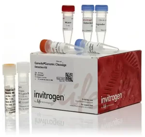
Invitrogen™GeneArt™ Genomic Cleavage Detection Kit
$440.54 Add to cart View Product DetailsThe GeneArt Genomic Cleavage Detection Kit provides a simple, reliable, and rapid method for the detection of locus-specific double-strand break formation. When using genome editing tools to obtain targeted mutations, it is necessary to determine the efficiency at which these nucleases cleave the target sequence, particularly prior to proceeding to the more laborious and expensive processes of cloning and sequencing.nnThe GeneArt genomic cleavage detection kit contains all of the reagents required prior to gel analysis. A sample of the edited cell population is used as a direct PCR template for amplification with primers specific to the targeted region. The PCR product is then denatured and re-annealed to produce heteroduplex mismatches where double strand breaks have occurred, resulting in indel introduction. These mismatches are recognized and cleaved by the detection enzyme. This cleavage is both easily detectable and quantifiable using gel analysis.
-
InvitrogenVEGF Monoclonal Antibody (16F1)
$575.00 Add to cart View Product DetailsnnThe VEGF-A monoclonal antibody clone # 16F1 (Product # M808) has successfully been paired as the coating antibody in a sandwich ELISA with detection antibody P802B (biotinylated conjugate of Product # P802).
Typical dilutions for sandwich ELISA include 1 µg/mL for coating and 0.125-0.5 µg/mL for detection.Target Information
VEGF (vascular endothelial growth factor) which is a 45 kDa homodimeric, disulfide-linked glycoprotein involved in angiogenesis which promotes tumor progression and metastasis. VEGF has a variety of effects on vascular endothelium, including the ability to promote endothelial cell viability, mitogenesis, chemotaxis, and vascular permeability. The VEGF family currently includes VEGF-A, VEGF-B, VEGF-C, VEGF-D, VEGF-E, and PIGF. VEGF and its receptor system have been shown to be the fundamental regulators in the cell signaling of angiogenesis. Most tumors have the absolute requirement of angiogenesis, and VEGF has been described as the most potent angiogenic cytokine linked to this process. To date 5 different isoforms of VEGF have been described. These isoforms are generated as the result of alternative splicing from a single VEGF gene. These various isoforms have been shown to bind to two tyrosine-kinase receptors flt-1 (VEGFR-1) and flk-1/KDR (VEGFR-2), which have been found to be expressed almost exclusively on endothelial cells. VEGF and its high-affinity binding receptors, the tyrosine kinases FLK1 and FLT1, are thought to be important for the development of embryonic vasculature. Studies have shown that an alternately spliced form of FLT1 produces a soluble protein, termed sFLT1, which binds vascular endothelial growth factor with high affinity, playing an inhibitory role in angiogenesis. Elevated levels of VEGF is linked to POEMS syndrome (Polyneuropathy, Organomegaly, Endocrinopathy, Monoclonal gammopathy, Skin changes) also known as Crow-Fukase syndrome which affects multiple organs in the body.
-
InvitrogenVEGF Monoclonal Antibody (JH121)
$506.00 Add to cart View Product DetailsMA5-13182 targets Vascular Endothelial Growth Factor in IF, WB, ICC, IHC, IP and ELISA applications and shows reactivity with Human and Rabbit samples.
nVEGF (vascular endothelial growth factor) which is a 45 kDa homodimeric, disulfide-linked glycoprotein involved in angiogenesis which promotes tumor progression and metastasis. VEGF has a variety of effects on vascular endothelium, including the ability to promote endothelial cell viability, mitogenesis, chemotaxis, and vascular permeability. The VEGF family currently includes VEGF-A, VEGF-B, VEGF-C, VEGF-D, VEGF-E, and PIGF. VEGF and its receptor system have been shown to be the fundamental regulators in the cell signaling of angiogenesis. Most tumors have the absolute requirement of angiogenesis, and VEGF has been described as the most potent angiogenic cytokine linked to this process. To date 5 different isoforms of VEGF have been described. These isoforms are generated as the result of alternative splicing from a single VEGF gene. These various isoforms have been shown to bind to two tyrosine-kinase receptors flt-1 (VEGFR-1) and flk-1/KDR (VEGFR-2), which have been found to be expressed almost exclusively on endothelial cells. VEGF and its high-affinity binding receptors, the tyrosine kinases FLK1 and FLT1, are thought to be important for the development of embryonic vasculature. Studies have shown that an alternately spliced form of FLT1 produces a soluble protein, termed sFLT1, which binds vascular endothelial growth factor with high affinity, playing an inhibitory role in angiogenesis. Elevated levels of VEGF is linked to POEMS syndrome (Polyneuropathy, Organomegaly, Endocrinopathy, Monoclonal gammopathy, Skin changes) also known as Crow-Fukase syndrome which affects multiple organs in the body.
-

InvitrogenVEGF Polyclonal Antibody
$512.90 Add to cart View Product DetailsThe VEGF-A monoclonal antibody clone No. 16F1 (Product No. M808) has successfully been paired as the coating antibody in a sandwich ELISA with detection antibody P802B (biotinylated conjugate of Product No. P802). Typical dilutions for sandwich ELISA include 1 µg/mL for coating and 0.125-0.5 µg/mL for detection.
nVEGF (vascular endothelial growth factor) which is a 45 kDa homodimeric, disulfide-linked glycoprotein involved in angiogenesis which promotes tumor progression and metastasis. VEGF has a variety of effects on vascular endothelium, including the ability to promote endothelial cell viability, mitogenesis, chemotaxis, and vascular permeability. The VEGF family currently includes VEGF-A, VEGF-B, VEGF-C, VEGF-D, VEGF-E, and PIGF. VEGF and its receptor system have been shown to be the fundamental regulators in the cell signaling of angiogenesis. Most tumors have the absolute requirement of angiogenesis, and VEGF has been described as the most potent angiogenic cytokine linked to this process. To date 5 different isoforms of VEGF have been described. These isoforms are generated as the result of alternative splicing from a single VEGF gene. These various isoforms have been shown to bind to two tyrosine-kinase receptors flt-1 (VEGFR-1) and flk-1/KDR (VEGFR-2), which have been found to be expressed almost exclusively on endothelial cells. VEGF and its high-affinity binding receptors, the tyrosine kinases FLK1 and FLT1, are thought to be important for the development of embryonic vasculature. Studies have shown that an alternately spliced form of FLT1 produces a soluble protein, termed sFLT1, which binds vascular endothelial growth factor with high affinity, playing an inhibitory role in angiogenesis. Elevated levels of VEGF is linked to POEMS syndrome (Polyneuropathy, Organomegaly, Endocrinopathy, Monoclonal gammopathy, Skin changes) also known as Crow-Fukase syndrome which affects multiple organs in the body.
-
InvitrogenVEGF Polyclonal Antibody, Biotin
$512.90 Add to cart View Product DetailsnnThe VEGF-A monoclonal antibody clone # 16F1 (Product # M808) has successfully been paired as the coating antibody in a sandwich ELISA with detection antibody P802B (biotinylated conjugate of Product # P802).
Typical dilutions for sandwich ELISA include 1 µg/mL for coating and 0.125-0.5 µg/mL for detection.Target Information
VEGF (vascular endothelial growth factor) which is a 45 kDa homodimeric, disulfide-linked glycoprotein involved in angiogenesis which promotes tumor progression and metastasis. VEGF has a variety of effects on vascular endothelium, including the ability to promote endothelial cell viability, mitogenesis, chemotaxis, and vascular permeability. The VEGF family currently includes VEGF-A, VEGF-B, VEGF-C, VEGF-D, VEGF-E, and PIGF. VEGF and its receptor system have been shown to be the fundamental regulators in the cell signaling of angiogenesis. Most tumors have the absolute requirement of angiogenesis, and VEGF has been described as the most potent angiogenic cytokine linked to this process. To date 5 different isoforms of VEGF have been described. These isoforms are generated as the result of alternative splicing from a single VEGF gene. These various isoforms have been shown to bind to two tyrosine-kinase receptors flt-1 (VEGFR-1) and flk-1/KDR (VEGFR-2), which have been found to be expressed almost exclusively on endothelial cells. VEGF and its high-affinity binding receptors, the tyrosine kinases FLK1 and FLT1, are thought to be important for the development of embryonic vasculature. Studies have shown that an alternately spliced form of FLT1 produces a soluble protein, termed sFLT1, which binds vascular endothelial growth factor with high affinity, playing an inhibitory role in angiogenesis. Elevated levels of VEGF is linked to POEMS syndrome (Polyneuropathy, Organomegaly, Endocrinopathy, Monoclonal gammopathy, Skin changes) also known as Crow-Fukase syndrome which affects multiple organs in the body.
-
InvitrogenVEGF Recombinant Polyclonal Antibody (1HCLC) X
$512.90 Add to cart View Product DetailsThis antibody is predicted to react with rat based on sequence homology.
Recombinant rabbit polyclonal antibodies are unique offerings from Thermo Fisher Scientific. They are comprised of a selection of multiple different recombinant monoclonal antibodies, providing the best of both worlds – the sensitivity of polyclonal antibodies with the specificity of monoclonal antibodies – all delivered with the consistency only found in a recombinant antibody. While functionally the same as a polyclonal antibody – recognizing multiple epitope sites on the target and producing higher detection sensitivity for low abundance targets – a recombinant rabbit polyclonal antibody has a known mixture of light and heavy chains. The exact population can be produced in every lot, circumventing the biological variability typically associated with polyclonal antibody production.Target Information
VEGF (vascular endothelial growth factor) which is a 45 kDa homodimeric, disulfide-linked glycoprotein involved in angiogenesis which promotes tumor progression and metastasis. VEGF has a variety of effects on vascular endothelium, including the ability to promote endothelial cell viability, mitogenesis, chemotaxis, and vascular permeability. The VEGF family currently includes VEGF-A, VEGF-B, VEGF-C, VEGF-D, VEGF-E, and PIGF. VEGF and its receptor system have been shown to be the fundamental regulators in the cell signaling of angiogenesis. Most tumors have the absolute requirement of angiogenesis, and VEGF has been described as the most potent angiogenic cytokine linked to this process. To date 5 different isoforms of VEGF have been described. These isoforms are generated as the result of alternative splicing from a single VEGF gene. These various isoforms have been shown to bind to two tyrosine-kinase receptors flt-1 (VEGFR-1) and flk-1/KDR (VEGFR-2), which have been found to be expressed almost exclusively on endothelial cells. VEGF and its high-affinity binding receptors, the tyrosine kinases FLK1 and FLT1, are thought to be important for the development of embryonic vasculature. Studies have shown that an alternately spliced form of FLT1 produces a soluble protein, termed sFLT1, which binds vascular endothelial growth factor with high affinity, playing an inhibitory role in angiogenesis. Elevated levels of VEGF is linked to POEMS syndrome (Polyneuropathy, Organomegaly, Endocrinopathy, Monoclonal gammopathy, Skin changes) also known as Crow-Fukase syndrome which affects multiple organs in the body.
-
InvitrogenVEGF-165 Polyclonal Antibody, Biotin, PeproTech®
$449.85 Add to cart View Product DetailsnnAA Sequence of recombinant protein: APMAEGGGQN HHEVVKFMDV YQRSYCHPIE TLVDIFQEYP DEIEYIFKPS CVPLMRCGGC CNDEGLECVP TEESNITMQI MRIKPHQGQH IGEMSFLQHN KCECRPKKDR ARQENPCGPC SERRKHLFVQ DPQTCKCSCK NTDSRCKARQ LELNERTCRC DKPRR.
Preparation: Produced from sera of rabbits immunized with highly pure Recombinant Human VEGF 165. Anti-Human VEGF 165-specific antibody was purified by affinity chromatography and then biotinylated.
Sandwich ELISA: To detect Human VEGF 165 by sandwich ELISA (using 100 µL/well antibody solution) a concentration of 0.25-1.0 µg/mL of this antibody is required. This biotinylated polyclonal antibody, in conjunction with PeproTech Polyclonal Anti-Human VEGF 165 (500-P10) as a capture antibody, allows the detection of at least 0.2-0.4 ng/well of Recombinant Human VEGF 165.
Western Blot: To detect hVEGF by Western Blot analysis this antibody can be used at a concentration of 0.1-0.2 µg/mL. Used in conjunction with compatible secondary reagents the detection limit for Recombinant hVEGF is 1.5-3.0 ng/lane, under either reducing or non-reducing conditions.Target Information
VEGF (vascular endothelial growth factor) which is a 45 kDa homodimeric, disulfide-linked glycoprotein involved in angiogenesis which promotes tumor progression and metastasis. VEGF has a variety of effects on vascular endothelium, including the ability to promote endothelial cell viability, mitogenesis, chemotaxis, and vascular permeability. The VEGF family currently includes VEGF-A, VEGF-B, VEGF-C, VEGF-D, VEGF-E, and PIGF. VEGF and its receptor system have been shown to be the fundamental regulators in the cell signaling of angiogenesis. Most tumors have the absolute requirement of angiogenesis, and VEGF has been described as the most potent angiogenic cytokine linked to this process. To date 5 different isoforms of VEGF have been described. These isoforms are generated as the result of alternative splicing from a single VEGF gene. These various isoforms have been shown to bind to two tyrosine-kinase receptors flt-1 (VEGFR-1) and flk-1/KDR (VEGFR-2), which have been found to be expressed almost exclusively on endothelial cells. VEGF and its high-affinity binding receptors, the tyrosine kinases FLK1 and FLT1, are thought to be important for the development of embryonic vasculature. Studies have shown that an alternately spliced form of FLT1 produces a soluble protein, termed sFLT1, which binds vascular endothelial growth factor with high affinity, playing an inhibitory role in angiogenesis. Elevated levels of VEGF is linked to POEMS syndrome (Polyneuropathy, Organomegaly, Endocrinopathy, Monoclonal gammopathy, Skin changes) also known as Crow-Fukase syndrome which affects multiple organs in the body.
-
InvitrogenVEGF-165 Polyclonal Antibody, Biotin, PeproTech®
$449.85 Add to cart View Product DetailsSequence of recombinant protein: APMAEGGGQN HHEVVKFMDV YQRSYCHPIE TLVDIFQEYP DEIEYIFKPS CVPLMRCGGC CNDEGLECVP TEESNITMQI MRIKPHQGQH IGEMSFLQHN KCECRPKKDR ARQENPCGPC SERRKHLFVQ DPQTCKCSCK NTDSRCKARQ LELNERTCRC DKPRR.
Preparation: Produced from sera of goats immunized with highly pure Recombinant Human VEGF 165. Anti-Human VEGF 165-specific antibody was purified by affinity chromatography and then biotinylated.
Sandwich ELISA: To detect Human VEGF 165 by sandwich ELISA (using 100 µL/well antibody solution) a concentration of 0.25-1.0 µg/mL of this antibody is required. This biotinylated polyclonal antibody, in conjunction with PeproTech Polyclonal Anti-Human VEGF 165 (500-P10G) as a capture antibody, allows the detection of at least 0.2-0.4 ng/well of Recombinant Human VEGF 165.
Western Blot: To detect hVEGF by Western Blot analysis this antibody can be used at a concentration of 0.1-0.2 µg/mL. Used in conjunction with compatible secondary reagents the detection limit for Recombinant hVEGF is 1.5-3.0 ng/lane, under either reducing or non-reducing conditions.Target Information
VEGF (vascular endothelial growth factor) which is a 45 kDa homodimeric, disulfide-linked glycoprotein involved in angiogenesis which promotes tumor progression and metastasis. VEGF has a variety of effects on vascular endothelium, including the ability to promote endothelial cell viability, mitogenesis, chemotaxis, and vascular permeability. The VEGF family currently includes VEGF-A, VEGF-B, VEGF-C, VEGF-D, VEGF-E, and PIGF. VEGF and its receptor system have been shown to be the fundamental regulators in the cell signaling of angiogenesis. Most tumors have the absolute requirement of angiogenesis, and VEGF has been described as the most potent angiogenic cytokine linked to this process. To date 5 different isoforms of VEGF have been described. These isoforms are generated as the result of alternative splicing from a single VEGF gene. These various isoforms have been shown to bind to two tyrosine-kinase receptors flt-1 (VEGFR-1) and flk-1/KDR (VEGFR-2), which have been found to be expressed almost exclusively on endothelial cells. VEGF and its high-affinity binding receptors, the tyrosine kinases FLK1 and FLT1, are thought to be important for the development of embryonic vasculature. Studies have shown that an alternately spliced form of FLT1 produces a soluble protein, termed sFLT1, which binds vascular endothelial growth factor with high affinity, playing an inhibitory role in angiogenesis. Elevated levels of VEGF is linked to POEMS syndrome (Polyneuropathy, Organomegaly, Endocrinopathy, Monoclonal gammopathy, Skin changes) also known as Crow-Fukase syndrome which affects multiple organs in the body.
-
InvitrogenVEGF-165 Polyclonal Antibody, Biotin, PeproTech®
$449.85 Add to cart View Product DetailsnnAA Sequence of recombinant protein: MAPTTEGEQK SHEVIKFMDV YQRSYCRPIE TLVDIFQEYP DEIEYIFKPS CVPLMRCAGC CNDEALECVP TSESNITMQI MRIKPHQSQH IGEMSFLQHS RCECRPKKDR TKPEKHCEPC SERRKHLFVQ DPQTCKCSCK NTDSRCKARQ LELNERTCRC DKPRR.
Preparation: Produced from sera of rabbits immunized with highly pure Recombinant Murine VEGF 165. Anti-Murine VEGF 165-specific antibody was purified by affinity chromatography and then biotinylated.
Sandwich ELISA: To detect Murine VEGF 165 by sandwich ELISA (using 100 µL/well antibody solution) a concentration of 0.25-1.0 µg/mL of this antibody is required. This biotinylated polyclonal antibody, in conjunction with PeproTech Polyclonal Anti-Murine VEGF 165 (500-P131) as a capture antibody, allows the detection of at least 0.2-0.4 ng/well of Recombinant Murine VEGF 165.
Western Blot: To detect mVEGF by Western Blot analysis this antibody can be used at a concentration of 0.1-0.2 µg/mL. Used in conjunction with compatible secondary reagents the detection limit for Recombinant mVEGF is 1.5-3.0 ng/lane, under either reducing or non-reducing conditions.Target Information
VEGF (vascular endothelial growth factor) which is a 45 kDa homodimeric, disulfide-linked glycoprotein involved in angiogenesis which promotes tumor progression and metastasis. VEGF has a variety of effects on vascular endothelium, including the ability to promote endothelial cell viability, mitogenesis, chemotaxis, and vascular permeability. The VEGF family currently includes VEGF-A, VEGF-B, VEGF-C, VEGF-D, VEGF-E, and PIGF. VEGF and its receptor system have been shown to be the fundamental regulators in the cell signaling of angiogenesis. Most tumors have the absolute requirement of angiogenesis, and VEGF has been described as the most potent angiogenic cytokine linked to this process. To date 5 different isoforms of VEGF have been described. These isoforms are generated as the result of alternative splicing from a single VEGF gene. These various isoforms have been shown to bind to two tyrosine-kinase receptors flt-1 (VEGFR-1) and flk-1/KDR (VEGFR-2), which have been found to be expressed almost exclusively on endothelial cells. VEGF and its high-affinity binding receptors, the tyrosine kinases FLK1 and FLT1, are thought to be important for the development of embryonic vasculature. Studies have shown that an alternately spliced form of FLT1 produces a soluble protein, termed sFLT1, which binds vascular endothelial growth factor with high affinity, playing an inhibitory role in angiogenesis. Elevated levels of VEGF is linked to POEMS syndrome (Polyneuropathy, Organomegaly, Endocrinopathy, Monoclonal gammopathy, Skin changes) also known as Crow-Fukase syndrome which affects multiple organs in the body.
-
InvitrogenVEGF-165 Polyclonal Antibody, Biotin, PeproTech®
$449.85 Add to cart View Product DetailsnnAA Sequence of recombinant protein: MAPTTEGEQK AHEVVKFMDV YQRSYCRPIE TLVDIFQEYP DEIEYIFKPS CVPLMRCAGC CNDEALECVP TSESNVTMQI MRIKPHQSQH IGEMSFLQHS RCECRPKKDR TKPEKHCEPC SERRKHLFVQ DPQTCKCSCK NTDSRCKARQ LELNERTCRC DKPRR.
Preparation: Produced from sera of rabbits immunized with highly pure Recombinant Rat VEGF 165. Anti-Rat VEGF 165-specific antibody was purified by affinity chromatography and then biotinylated.
Sandwich ELISA: To detect Rat VEGF by sandwich ELISA (using 100 µL/well antibody solution) a concentration of 0.25-1.0 µg/mL of this antibody is required. This biotinylated polyclonal antibody, in conjunction with PeproTech Polyclonal Anti-Rat VEGF (500-P275) as a capture antibody, allows the detection of at least 0.2-0.4 ng/well of Recombinant Rat VEGF.
Western Blot: To detect Rat VEGF by Western Blot analysis this antibody can be used at a concentration of 0.1-0.2 µg/mL. Used in conjunction with compatible secondary reagents the detection limit for Recombinant Rat VEGF is 1.5-3.0 ng/lane, under either reducing or non-reducing conditions.Target Information
VEGF (vascular endothelial growth factor) which is a 45 kDa homodimeric, disulfide-linked glycoprotein involved in angiogenesis which promotes tumor progression and metastasis. VEGF has a variety of effects on vascular endothelium, including the ability to promote endothelial cell viability, mitogenesis, chemotaxis, and vascular permeability. The VEGF family currently includes VEGF-A, VEGF-B, VEGF-C, VEGF-D, VEGF-E, and PIGF. VEGF and its receptor system have been shown to be the fundamental regulators in the cell signaling of angiogenesis. Most tumors have the absolute requirement of angiogenesis, and VEGF has been described as the most potent angiogenic cytokine linked to this process. To date 5 different isoforms of VEGF have been described. These isoforms are generated as the result of alternative splicing from a single VEGF gene. These various isoforms have been shown to bind to two tyrosine-kinase receptors flt-1 (VEGFR-1) and flk-1/KDR (VEGFR-2), which have been found to be expressed almost exclusively on endothelial cells. VEGF and its high-affinity binding receptors, the tyrosine kinases FLK1 and FLT1, are thought to be important for the development of embryonic vasculature. Studies have shown that an alternately spliced form of FLT1 produces a soluble protein, termed sFLT1, which binds vascular endothelial growth factor with high affinity, playing an inhibitory role in angiogenesis. Elevated levels of VEGF is linked to POEMS syndrome (Polyneuropathy, Organomegaly, Endocrinopathy, Monoclonal gammopathy, Skin changes) also known as Crow-Fukase syndrome which affects multiple organs in the body.
-
InvitrogenVEGF-165 Polyclonal Antibody, PeproTech®
$366.95 Add to cart View Product DetailsnnAA Sequence of recombinant protein: APMAEGGGQN HHEVVKFMDV YQRSYCHPIE TLVDIFQEYP DEIEYIFKPS CVPLMRCGGC CNDEGLECVP TEESNITMQI MRIKPHQGQH IGEMSFLQHN KCECRPKKDR ARQENPCGPC SERRKHLFVQ DPQTCKCSCK NTDSRCKARQ LELNERTCRC DKPRR.
Preparation: Produced from sera of rabbits immunized with highly pure Recombinant Human VEGF 165. Anti-Human VEGF 165-specific antibody was purified by affinity chromatography employing an immobilized Human VEGF 165 matrix.
Sandwich ELISA: To detect Human VEGF 165 by sandwich ELISA (using 100 µL/well antibody solution) a concentration of 0.5-2.0 µg/mL of this antibody is required. This antigen affinity purified antibody, in conjunction with PeproTech Biotinylated Anti-Human VEGF 165 (500-P10Bt) as a detection antibody, allows the detection of at least 0.2-0.4 ng/well of Recombinant Human VEGF 165.
Western Blot: To detect Human VEGF 165 by Western Blot analysis this antibody can be used at a concentration of 0.1-0.2 µg/mL. Used in conjunction with compatible secondary reagents the detection limit for Recombinant Human VEGF 165 is 1.5-3.0 ng/lane, under either reducing or non-reducing conditions.Target Information
VEGF (vascular endothelial growth factor) which is a 45 kDa homodimeric, disulfide-linked glycoprotein involved in angiogenesis which promotes tumor progression and metastasis. VEGF has a variety of effects on vascular endothelium, including the ability to promote endothelial cell viability, mitogenesis, chemotaxis, and vascular permeability. The VEGF family currently includes VEGF-A, VEGF-B, VEGF-C, VEGF-D, VEGF-E, and PIGF. VEGF and its receptor system have been shown to be the fundamental regulators in the cell signaling of angiogenesis. Most tumors have the absolute requirement of angiogenesis, and VEGF has been described as the most potent angiogenic cytokine linked to this process. To date 5 different isoforms of VEGF have been described. These isoforms are generated as the result of alternative splicing from a single VEGF gene. These various isoforms have been shown to bind to two tyrosine-kinase receptors flt-1 (VEGFR-1) and flk-1/KDR (VEGFR-2), which have been found to be expressed almost exclusively on endothelial cells. VEGF and its high-affinity binding receptors, the tyrosine kinases FLK1 and FLT1, are thought to be important for the development of embryonic vasculature. Studies have shown that an alternately spliced form of FLT1 produces a soluble protein, termed sFLT1, which binds vascular endothelial growth factor with high affinity, playing an inhibitory role in angiogenesis. Elevated levels of VEGF is linked to POEMS syndrome (Polyneuropathy, Organomegaly, Endocrinopathy, Monoclonal gammopathy, Skin changes) also known as Crow-Fukase syndrome which affects multiple organs in the body.
-
InvitrogenVEGF-165 Polyclonal Antibody, PeproTech®
$366.95 Add to cart View Product DetailsnnAA Sequence of recombinant protein: APMAEGGGQN HHEVVKFMDV YQRSYCHPIE TLVDIFQEYP DEIEYIFKPS CVPLMRCGGC CNDEGLECVP TEESNITMQI MRIKPHQGQH IGEMSFLQHN KCECRPKKDR ARQENPCGPC SERRKHLFVQ DPQTCKCSCK NTDSRCKARQ LELNERTCRC DKPRR.
Preparation: Produced from sera of goats immunized with highly pure Recombinant Human VEGF 165. Anti-Human VEGF 165-specific antibody was purified by affinity chromatography employing an immobilized Human VEGF 165 matrix.
Sandwich ELISA: To detect Human VEGF 165 by sandwich ELISA (using 100 µL/well antibody solution) a concentration of 0.5-2.0 µg/mL of this antibody is required. This antigen affinity purified antibody, in conjunction with PeproTech Biotinylated Anti-Human VEGF 165 (500-P10GBt) as a detection antibody, allows the detection of at least 0.2-0.4 ng/well of Recombinant Human VEGF 165.
Western Blot: To detect Human VEGF 165 by Western Blot analysis this antibody can be used at a concentration of 0.1-0.2 µg/mL. Used in conjunction with compatible secondary reagents the detection limit for Recombinant Human VEGF 165 is 1.5-3.0 ng/lane, under either reducing or non-reducing conditions.Target Information
VEGF (vascular endothelial growth factor) which is a 45 kDa homodimeric, disulfide-linked glycoprotein involved in angiogenesis which promotes tumor progression and metastasis. VEGF has a variety of effects on vascular endothelium, including the ability to promote endothelial cell viability, mitogenesis, chemotaxis, and vascular permeability. The VEGF family currently includes VEGF-A, VEGF-B, VEGF-C, VEGF-D, VEGF-E, and PIGF. VEGF and its receptor system have been shown to be the fundamental regulators in the cell signaling of angiogenesis. Most tumors have the absolute requirement of angiogenesis, and VEGF has been described as the most potent angiogenic cytokine linked to this process. To date 5 different isoforms of VEGF have been described. These isoforms are generated as the result of alternative splicing from a single VEGF gene. These various isoforms have been shown to bind to two tyrosine-kinase receptors flt-1 (VEGFR-1) and flk-1/KDR (VEGFR-2), which have been found to be expressed almost exclusively on endothelial cells. VEGF and its high-affinity binding receptors, the tyrosine kinases FLK1 and FLT1, are thought to be important for the development of embryonic vasculature. Studies have shown that an alternately spliced form of FLT1 produces a soluble protein, termed sFLT1, which binds vascular endothelial growth factor with high affinity, playing an inhibitory role in angiogenesis. Elevated levels of VEGF is linked to POEMS syndrome (Polyneuropathy, Organomegaly, Endocrinopathy, Monoclonal gammopathy, Skin changes) also known as Crow-Fukase syndrome which affects multiple organs in the body.
-
InvitrogenVEGF-165 Polyclonal Antibody, PeproTech®
$381.01 Add to cart View Product DetailsnnAA Sequence of recombinant protein: MAPTTEGEQK SHEVIKFMDV YQRSYCRPIE TLVDIFQEYP DEIEYIFKPS CVPLMRCAGC CNDEALECVP TSESNITMQI MRIKPHQSQH IGEMSFLQHS RCECRPKKDR TKPEKHCEPC SERRKHLFVQ DPQTCKCSCK NTDSRCKARQ LELNERTCRC DKPRR.
Preparation: Produced from sera of rabbits immunized with highly pure Recombinant Murine VEGF 165. Anti-Murine VEGF 165-specific antibody was purified by affinity chromatography employing an immobilized Murine VEGF 165 matrix.
Sandwich ELISA: To detect Murine VEGF 165 by sandwich ELISA (using 100 µL/well antibody solution) a concentration of 0.5-2.0 µg/mL of this antibody is required. This antigen affinity purified antibody, in conjunction with PeproTech Biotinylated Anti-Murine VEGF 165 (500-P131Bt) as a detection antibody, allows the detection of at least 0.2-0.4 ng/well of Recombinant Murine VEGF 165.
Western Blot: To detect Murine VEGF 165 by Western Blot analysis this antibody can be used at a concentration of 0.1-0.2 µg/mL. Used in conjunction with compatible secondary reagents the detection limit for Recombinant Murine VEGF165 is 1.5-3.0 ng/lane, under either reducing or non-reducing conditions.Target Information
VEGF (vascular endothelial growth factor) which is a 45 kDa homodimeric, disulfide-linked glycoprotein involved in angiogenesis which promotes tumor progression and metastasis. VEGF has a variety of effects on vascular endothelium, including the ability to promote endothelial cell viability, mitogenesis, chemotaxis, and vascular permeability. The VEGF family currently includes VEGF-A, VEGF-B, VEGF-C, VEGF-D, VEGF-E, and PIGF. VEGF and its receptor system have been shown to be the fundamental regulators in the cell signaling of angiogenesis. Most tumors have the absolute requirement of angiogenesis, and VEGF has been described as the most potent angiogenic cytokine linked to this process. To date 5 different isoforms of VEGF have been described. These isoforms are generated as the result of alternative splicing from a single VEGF gene. These various isoforms have been shown to bind to two tyrosine-kinase receptors flt-1 (VEGFR-1) and flk-1/KDR (VEGFR-2), which have been found to be expressed almost exclusively on endothelial cells. VEGF and its high-affinity binding receptors, the tyrosine kinases FLK1 and FLT1, are thought to be important for the development of embryonic vasculature. Studies have shown that an alternately spliced form of FLT1 produces a soluble protein, termed sFLT1, which binds vascular endothelial growth factor with high affinity, playing an inhibitory role in angiogenesis. Elevated levels of VEGF is linked to POEMS syndrome (Polyneuropathy, Organomegaly, Endocrinopathy, Monoclonal gammopathy, Skin changes) also known as Crow-Fukase syndrome which affects multiple organs in the body.
-
InvitrogenVEGF-165 Polyclonal Antibody, PeproTech®
$351.69 Add to cart View Product DetailsProduct Specific Information
Sequence of recombinant protein: MAPTTEGEQK AHEVVKFMDV YQRSYCRPIE TLVDIFQEYP DEIEYIFKPS CVPLMRCAGC CNDEALECVP TSESNVTMQI MRIKPHQSQH IGEMSFLQHS RCECRPKKDR TKPEKHCEPC SERRKHLFVQ DPQTCKCSCK NTDSRCKARQ LELNERTCRC DKPRR.
Preparation: Produced from sera of rabbits immunized with highly pure Recombinant Rat VEGF 165. Anti-Rat VEGF 165-specific antibody was purified by affinity chromatography employing an immobilized Rat VEGF 165 matrix.
Sandwich ELISA: To detect Rat VEGF 165 by sandwich ELISA (using 100 µL/well antibody solution) a concentration of 0.5-2.0 µg/mL of this antibody is required. This antigen affinity purified antibody, in conjunction with PeproTech Biotinylated Anti-Rat VEGF 165 (500-P275Bt) as a detection antibody, allows the detection of at least 0.2-0.4 ng/well of Recombinant Rat VEGF 165.
Western Blot: To detect Rat VEGF 165 by Western Blot analysis this antibody can be used at a concentration of 0.1-0.2 µg/mL. Used in conjunction with compatible secondary reagents the detection limit for Recombinant Rat VEGF 165 is 1.5-3.0 ng/lane, under either reducing or non-reducing conditions.Target Information
VEGF (vascular endothelial growth factor) which is a 45 kDa homodimeric, disulfide-linked glycoprotein involved in angiogenesis which promotes tumor progression and metastasis. VEGF has a variety of effects on vascular endothelium, including the ability to promote endothelial cell viability, mitogenesis, chemotaxis, and vascular permeability. The VEGF family currently includes VEGF-A, VEGF-B, VEGF-C, VEGF-D, VEGF-E, and PIGF. VEGF and its receptor system have been shown to be the fundamental regulators in the cell signaling of angiogenesis. Most tumors have the absolute requirement of angiogenesis, and VEGF has been described as the most potent angiogenic cytokine linked to this process. To date 5 different isoforms of VEGF have been described. These isoforms are generated as the result of alternative splicing from a single VEGF gene. These various isoforms have been shown to bind to two tyrosine-kinase receptors flt-1 (VEGFR-1) and flk-1/KDR (VEGFR-2), which have been found to be expressed almost exclusively on endothelial cells. VEGF and its high-affinity binding receptors, the tyrosine kinases FLK1 and FLT1, are thought to be important for the development of embryonic vasculature. Studies have shown that an alternately spliced form of FLT1 produces a soluble protein, termed sFLT1, which binds vascular endothelial growth factor with high affinity, playing an inhibitory role in angiogenesis. Elevated levels of VEGF is linked to POEMS syndrome (Polyneuropathy, Organomegaly, Endocrinopathy, Monoclonal gammopathy, Skin changes) also known as Crow-Fukase syndrome which affects multiple organs in the body.
-
J Chain Monoclonal Antibody (SP105), Invitrogen™
$667.00 Add to cart View Product DetailsHeat-mediated antigen retrieval is recommended prior to staining, using a 10mM citrate buffer, pH 6.0, for 10 minutes followed by cooling at room temperature for 20 min. Following antigen retrieval, incubate samples with primary antibody for 10 min at room temperature. A suggested positive control is tonsil tissue.
nJ chain is a small glycopeptide and is structurally unrelated to heavy or light chains, but is synthesized by all plasma cells that secrete polymeric immunoglobulins. J chain is linked to IgA and IgM by disulfide bonds, however, it has been detected in IgG- and IgD-containing cells. J chains are present in a large proportion of the immunoglobulin-positive cells in the germinal centers of the tonsil and lymph node. B cells secrete J chain at an early stage of differentiation with the expression persisting in those cells destined to produce IgA or IgM.
-
LargeFlex adapter 2 x 250 mL tube holder on FastPrep-96
$2,986.76 Add to cart View Product Details2 x 250 mL tube holder for room temperature grinding on FastPrep-96.
2,597.18 -
Lipofectamine™ 3000 Transfection Reagent
$753.73 Add to cart View Product DetailsLipofectamine™ 3000 Transfection Reagent leverages our most advanced lipid nanoparticle technology to provide superior transfection performance with improved application outcomes and reproducible results.
nThis reagent delivers superior transfection efficiency and improved cell viability for the widest range of hard-to-transfect and common cells (e.g., HEK293, HeLa).
Successfully transfect the widest variety of biologically relevant cell types with a reagent that offers:-
- Superior performance—our highest efficiency reagent for difficult-to-transfect cells
n
-
- Improved cell viability—gentle on your cells, with low toxicity
n
-
- Versatile—one reagent for DNA, RNA, and co-transfection
n
-
-
LIVE/DEAD™ Fixable Aqua Dead Cell Stain Kit, for 405 nm excitation
$454.25 Add to cart View Product Details- Used to determine the viability of cells prior to the fixation and permeabilization required for intracellular antibody staining or prior to elimination of biohazardous materials using formaldehyde fixation
- Kit has been optimized and validated for use with a violet laser flow cytometer
- Conveniently packaged in 40-test vials to help ensure the stability and performance of the dye over time
- Amine reactive dyes in solution will lose their effectiveness over a short period of time, therefore it is recommended to completely use the vial once rehydrated
- If this is not possible, aliquot the vials in small volumes and store at -80°C, avoiding freeze-thaw cycles
- LIVE/DEAD™ Fixable Dead Cell Stains are available in a wide variety of colors to meet multi-color panel needs
Low compensation
- The LIVE/DEAD™ Fixable Aqua Stain was selected based on its fluorescent properties to minimize compensation between other violet dyes and dyes that excite off of the 488 nm blue laser
- The aqua-fluorescent reactive dye has an excitation maximum of ?375 nm, but it is excited well with the 405 nm violet laser
- It has an emission maxima of ?512 nm, so it can be collected in the second channel on most violet laser flow cytometers
-
LIVE/DEAD™ Fixable Near-IR Dead Cell Stain Kit, for 633 or 635 nm excitation
$529.28 Add to cart View Product Details-
- Used to determine the viability of cells prior to the fixation and permeabilization required for intracellular antibody staining or prior to elimination of biohazardous materials using formaldehyde fixation
n
-
- Kit has been optimized and validated for use with a violet laser flow cytometer
n
-
- Conveniently packaged in 40-test vials to help ensure the stability and performance of the dye over time
n
-
- Amine reactive dyes in solution will lose their effectiveness over a short period of time, therefore it is recommended to completely use the vial once rehydrated
n
-
- If this is not possible, aliquot the vials in small volumes and store at -80°C, avoiding freeze-thaw cycles
n
-
- LIVE/DEAD™ Fixable Dead Cell Stains are available in a wide variety of colors to meet multi-color panel needs
n
-
-
Lock pins for dome locking
$33.94 Add to cart View Product DetailsReplacement locking pins for FastPrep-24 Classic dome.
-
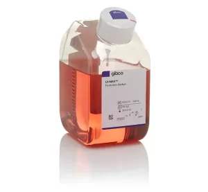
LV-MAX™ Production Medium
$361.10 Add to cart View Product DetailsGibco LV-MAX Production Medium is a chemically defined, serum-free, protein-free medium, specially developed for high-density growth and transfection of Gibco Viral Production Cells (suspension-adapted HEK 293-derived cells) promoting high-titer lentiviral production.
-
- Supports growth of suspension Viral Production Cells to densities of over 1.0 x 10⁷ cells/mL
n
-
- Enables sustained, high-level production of high-density transiently transfected cultures, achieving titer of up to 1 x 10⁸ TU/mL (unconcentrated LVVs-GFP)
n
-
- Supports growth and transfection of Viral Production Cells in culture formats from 96-deep well blocks to 2-L flasks and bioreactors
n
-
-
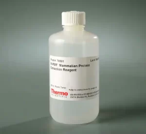
M-PER™ Mammalian Protein Extraction Reagent
$441.60 Add to cart View Product DetailsThermo Scientific™ M-PER Mammalian Protein Extraction Reagent is designed to provide highly efficient total protein extraction from cultured mammalian cells.
nThe complete cell lysis reagent is a nondenaturing detergent formulation that dissolves cell membranes and extracts total soluble cellular protein in only 5 minutes. M-PER Reagent requires little or no mechanical disruption and yields more protein than freeze/thaw cycles and sonication. This mammalian cell lysis reagent is so efficient that adherent cells do not need to be scraped from the culture dish, enabling easy and direct lysis and analysis of cells grown in 24-well and 96-well plates. Resulting cell lysates from either adherent or suspension cells are compatible with many downstream assays including immunoassays, enzyme assays and a variety of common reporter assays. Highlights:
-
- Gentle – mild detergent lysis, yielding extracts that are immediately compatible with Coomassie™ (Bradford) and BCA protein assays or SDS-PAGE
n
-
- Compatible – extracts soluble proteins in nondenatured state, enabling direct use in immunoprecipitation and other affinity purification procedures
n
-
- Amine-free and dialyzable – formulation ensures compatibility with subsequent assay systems
n
-
- Convenient – lyse adherent cells directly in plate or after scraping and washing in suspension
n
-
- Non-denaturing – maintain luciferase, ß-galactosidase, CAT and other reporter gene activities as well or better than other suppliers’ products and freeze/thaw methods
n
-
-
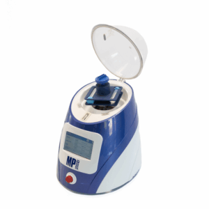
MagFlex-96 aNAP System RUO
$30,092.63 Add to cart View Product DetailsMagFlex-96 aNAP System RUO
26,167.50 -
Melatonin ELISA kit
$1,366.48 Add to cart View Product DetailsMelatonin ELISA kit
1,188.24 -
Metal BigPrep™ adapter for 2×50 mL tube holder on FastPrep24
$2,527.69 Add to cart View Product DetailsAdapter for FastPrep-24 instrument. Holds 2 x 50 mL metal tubes. To grind or homogenize most difficult samples. Can resist high temperatures up to 450°C
2,197.99 -
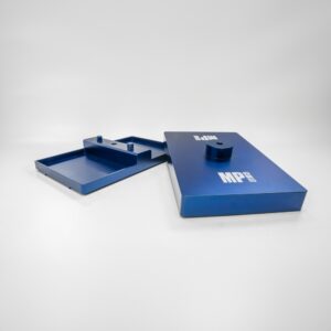
Metal Dual Plate Holder Assembly for FastPrep-96
$3,448.28 Add to cart View Product Details-
Adapter for FastPrep-96 instrument
-
Holds 2 x 96 deep well plates
-
Anodized aluminum metal
-
Wide temperature range resistance
2,998.50 -
-
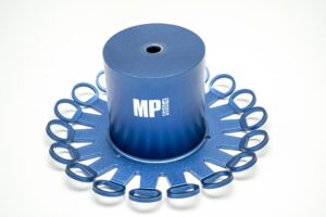
Metal MidiPrep Adapter for 18 x 5 mL for FastPrep-24
$1,292.03 Add to cart View Product Details- Adapter for FastPrep-24 5G and Classic
- Holds 18 x 5 mL tubes
- Grind or homogenize most difficult samples
- Can resist high temperatures up to 450°C
1,123.50 -
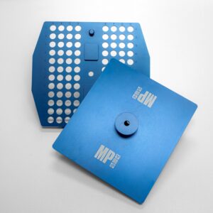
Metal QuickFlex Adapter for 96 x 2 mL tubes on FastPrep-96
$2,672.03 Add to cart View Product DetailsMetal QuickFlex Adapter for 96 x 2 mL tubes on FastPrep-96
2,323.50